
Female Hormone Testing Panel Essentials to Uncover Subtle Imbalances
What Is Included in a Female Hormone Testing Panel?
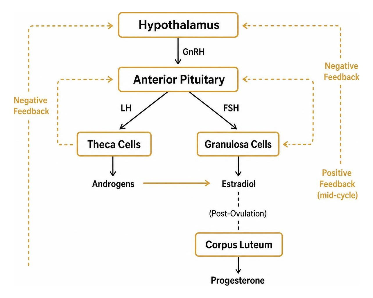
A female hormone testing panel includes key reproductive sex steroids such as estradiol, progesterone, and testosterone, pituitary gonadotropins (LH and FSH), adrenal androgens like DHEA-S, carrier proteins such as SHBG, and thyroid and metabolic markers including TSH, free T4, and free T3. Together, these biomarkers reveal how the hypothalamus, pituitary gland, ovaries, thyroid, and adrenal glands are communicating.
The female endocrine network relies on intricate feedback loops between these organ systems. When one hormone drifts out of range, downstream systems adapt, often masking the root cause of fatigue, weight fluctuations, or menstrual disruption.
A thorough assessment measures the production of primary sex steroids along with the signaling proteins that control them.
Hormone Biomarker | Primary Origin | Core Physiological Role | Key Clinical Indicators When Out of Range |
|---|---|---|---|
Estradiol (E2) | Ovaries | Regulates menstrual cycle, bone density, vascular elasticity, and mood | Low: Hot flashes, vaginal dryness, bone loss. High: Heavy menses, breast tenderness |
Progesterone | Corpus Luteum | Stabilizes the endometrium, supports sleep quality, and balances estrogen | Low: Short luteal phase, spotting, anxiety, insomnia |
Luteinizing Hormone (LH) | Pituitary Gland | Triggers ovulation; stimulates ovarian theca cells | Elevated LH:FSH ratio: Ovulatory dysfunction, polycystic ovarian patterns |
Follicle-Stimulating Hormone (FSH) | Pituitary Gland | Recruits and matures ovarian follicles | Elevated: Diminished ovarian reserve, perimenopause, menopause |
Total & Free Testosterone | Ovaries & Adrenals | Maintains lean muscle mass, bone strength, libido, and energy | Low: Muscle loss, poor recovery, low drive. High: Hirsutism, cystic acne, cycle loss |
DHEA-S | Adrenal Glands | Androgen precursor; supports cellular vitality and stress adaptation | Low: Adrenal exhaustion, fatigue. High: Androgen excess, stress response |
Sex Hormone-Binding Globulin (SHBG) | Liver | Transports sex steroids; regulates free, bioavailable hormone fractions | Low: High free androgens, insulin resistance. High: Low free sex steroids |
Thyroid-Stimulating Hormone (TSH) | Pituitary Gland | Regulates thyroid production; master metabolic regulator | High: Hypothyroidism, fatigue, weight gain. Low: Hyperthyroidism |
Key Reproductive and Adrenal Hormones Measured
Evaluating ovarian function requires looking at both estradiol and progesterone. Estradiol builds the uterine lining and supports joint, cognitive, and cardiovascular health. Progesterone counterbalances estrogenic stimulation, keeping mood steady and promoting deep restorative sleep. Testing both helps assess the ovulatory cycle and identifies whether luteal phase insufficiency is contributing to premenstrual tension or mid-cycle spotting.
We also examine pituitary gonadotropins—luteinizing hormone (LH) and follicle-stimulating hormone (FSH). Tracking the relationship between LH and FSH sheds light on follicular development. A baseline assessment of total testosterone, free testosterone, and DHEA-S (dehydroepiandrosterone sulfate) highlights androgen balance. Women need testosterone for bone mineral density, neuromuscular drive, and metabolic flexibility.
Because circulating hormones bind to proteins, measuring Sex Hormone-Binding Globulin (SHBG) is essential to calculate your free androgen index. Tracking these hormones gives clear context to our work analyzing performance biomarkers in active individuals.
Thyroid and Metabolic Markers for Comprehensive Insight
Reproductive health does not operate in isolation from metabolic health. Thyroid disorders are significantly more common in women than men, making thyroid-stimulating hormone (TSH), free thyroxine (Free T4), and free triiodothyronine (Free T3) central to any female blood panel. Thyroid autoimmunity, driven by thyroid peroxidase (TPO) antibodies, can disrupt menstrual regularity and cellular energy years before standard TSH levels trigger a clinical flag.
Prolactin is another key pituitary hormone; mild elevations can suppress GnRH pulsation, causing missed periods or unexplained infertility. Evaluating fasting insulin alongside these endocrine markers is equally vital. Insulin resistance directly stimulates ovarian theca cells to overproduce androgens, while lowering SHBG in the liver. Incorporating broader metabolic screens alongside sex steroids lets us assess cardiovascular risk and systemic stress while evaluating inflammation blood markers.
Signs and Symptoms Indicating You Need Hormonal Testing
When hormone production falls out of balance, the body provides subtle clues before severe symptoms develop. Many women spend months or years attributing brain fog, low energy, and sluggish recovery to poor sleep or everyday stress. Testing replaces guesswork with measurable data.
Recognizing Menstrual, Mood, and Energy Shifts
Cycle irregularities are among the clearest signs of an underlying imbalance. These can present as:
Amenorrhea: Missing periods for three consecutive months or more.
Oligomenorrhea: Extended cycles lasting longer than 35 days.
Luteal Phase Defects: Short cycles with spotting beginning 4 to 6 days before full menses, signaling low progesterone.
Severe PMS or PMDD: Marked mood swings, fluid retention, migraines, and intense irritability.
Energy and Cognitive Changes: Waking up unrefreshed, enduring mid-afternoon energy crashes, and dealing with chronic brain fog.
Athletic Plateaus: Slower recovery, unexpected strength loss, persistent joint discomfort, or an elevated resting heart rate despite structured training.
Screening for PCOS, Perimenopause, and Thyroid Dysfunction
Polycystic ovary syndrome (PCOS) affects an estimated 6–12% of women of reproductive age (with broader diagnostic criteria identifying 8–13%). Between 50% and 70% of women with PCOS exhibit some degree of insulin resistance. Classic signs include cystic acne along the jawline, hirsutism (excess facial or body hair), and stubborn visceral weight gain driven by elevated androgens.
At the other end of the reproductive spectrum, perimenopause often begins in a woman's late 30s or early 40s. The transition is marked by wide swings in estradiol, hot flashes, night sweats, sleep fragmentation, and unexpected cycle variations.
Concurrently, subclinical hypothyroidism or Hashimoto’s thyroiditis can mirror these symptoms, causing cold intolerance, thinning hair, dry skin, and constipation. Standardized panels help untangle these overlapping presentations so you can pursue targeted protocols.
Timing Rules and Preparation for Accurate Results
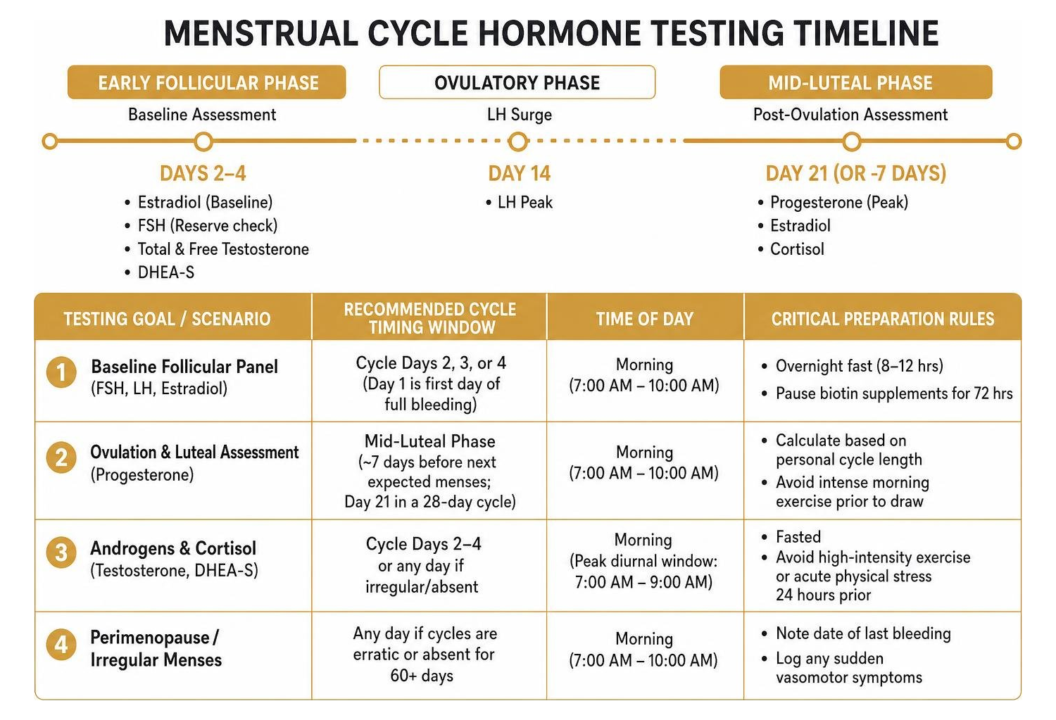
A hormone test is only as good as the conditions under which it was collected. Because female sex hormones rise and fall across the menstrual cycle and follow daily circadian patterns, timing errors can yield misleading data.
Testing Goal / Scenario | Recommended Cycle Timing Window | Time of Day | Critical Preparation Rules |
|---|---|---|---|
Baseline Follicular Panel (FSH, LH, Estradiol) | Cycle Days 2, 3, or 4 (Day 1 is first day of full bleeding) | Morning (7:00 AM – 10:00 AM) | Overnight fast (8–12 hrs); pause biotin supplements for 72 hrs |
Ovulation & Luteal Assessment (Progesterone) | Mid-Luteal Phase (~7 days before next expected menses; Day 21 in a 28-day cycle) | Morning (7:00 AM – 10:00 AM) | Calculate based on personal cycle length; avoid intense morning exercise prior to draw |
Androgens & Cortisol (Testosterone, DHEA-S) | Cycle Days 2–4 or any day if irregular/absent | Morning (Peak diurnal window: 7:00 AM – 9:00 AM) | Fasted; avoid high-intensity exercise or acute physical stress 24 hours prior |
Perimenopause / Irregular Menses | Any day if cycles are erratic or absent for 60+ days | Morning (7:00 AM – 10:00 AM) | Note date of last bleeding; log any sudden vasomotor symptoms |
Menstrual Cycle Timing: Follicular vs. Luteal Phase
To measure baseline ovarian signaling, schedule blood draws during the early follicular phase (Cycle Days 2 to 4). During this window, estrogen and progesterone are at their lowest baseline levels, allowing clinicians to evaluate whether pituitary signaling (FSH and LH) is balanced and whether estradiol output matches your reproductive stage.
Testing progesterone requires a different approach. Progesterone peaks during the mid-luteal phase, after the follicle releases an egg and transforms into the corpus luteum. The common convention of drawing on "Day 21" assumes a textbook 28-day cycle with ovulation occurring precisely on Day 14.
If your cycle runs 32 days, ovulation typically occurs around Day 18, meaning a Day 21 draw would capture progesterone before its peak, falsely suggesting anovulation. Instead, aim to test 7 days before your anticipated period. For women with absent or completely unpredictable cycles, testing can be done on any day to establish a baseline.
Avoiding Testing Interference from Biotin and Medications
Preparation before sample collection helps protect data integrity:
Biotin Washout: High-dose biotin (vitamin B7), often found in hair, skin, and nail supplements or multivitamins, directly interferes with streptavidin-biotin immunoassays used by modern clinical laboratories. This can falsely elevate free thyroid hormones and estradiol while falsely depressing TSH. Stop taking supplemental biotin for at least 72 hours before blood collection.
Contraceptives and HRT: Combined oral contraceptive pills, progestin-only pills, patches, and hormonal IUDs suppress endogenous hypothalamic-pituitary-ovarian signaling. While testing on hormonal contraception shows the systemic impact of those exogenous hormones, it will not reflect your natural baseline production.
Fasting and Circadian Controls: Fast for 8 to 12 hours before your blood draw, drinking only water. Collect blood in the early morning between 7:00 AM and 10:00 AM, when circulating concentrations of testosterone, DHEA-S, and cortisol are at their natural peak.
Comparing Specimen Types and Testing Methods
Navigating testing options means weighing convenience against analytical reliability across different specimen types.

Blood Serum, Saliva, and Dried Urine Testing
Venipuncture Serum Blood (Gold Standard): Clinical blood draws analyzed via liquid chromatography-tandem mass spectrometry (LC/MS-MS) or automated chemiluminescent immunoassay provide high clinical accuracy. Blood testing is the universal benchmark used by endocrinologists to assess total concentrations of circulating sex steroids, thyroid hormones, and pituitary signaling markers.
Salivary Testing: Saliva captures unbound, free steroid fractions that pass through salivary glands. It is non-invasive and helpful for mapping the diurnal cortisol awakening response across multiple time points. However, salivary steroid levels can vary significantly based on oral microbiome health, gum health, and local sample collection technique.
Dried Urine Testing: Dried urine testing measures downstream hormone metabolites over 24 hours. This reveals how your liver clears estrogen into 2-hydroxy, 4-hydroxy, or 16-hydroxy forms and provides insight into overall adrenal metabolite production.
Capillary Fingerstick Kits: While convenient for mail-in testing, capillary blood collection can suffer from hemolysis or insufficient sample volume if the finger is squeezed excessively during collection, which can alter sensitive peptide readings.
Choosing the Right Female Hormone Testing Panel: Basic vs. Expanded
Standard consumer panels vary in scope and depth. Basic direct-access panels provide an affordable entry point for reproductive health screening:
Basic Panels ($150 to $225): Typically measure 6 to 8 core biomarkers: Estradiol, Progesterone, Total Testosterone, LH, FSH, DHEA-S, and TSH. These panels are well-suited for a straightforward baseline check or initial fertility and cycle-regularity screens.
Expanded Panels ($289 to $450+): Add critical layers including Free Testosterone, SHBG, Free T3, Free T4, TPO Antibodies, Prolactin, Cortisol, and sometimes high-sensitivity CRP.
Selecting an expanded profile provides the detailed endocrine context needed to inform biomarker-based nutrition plans and adjust athletic periodization.
Navigating Results, Reference Ranges, and Next Steps
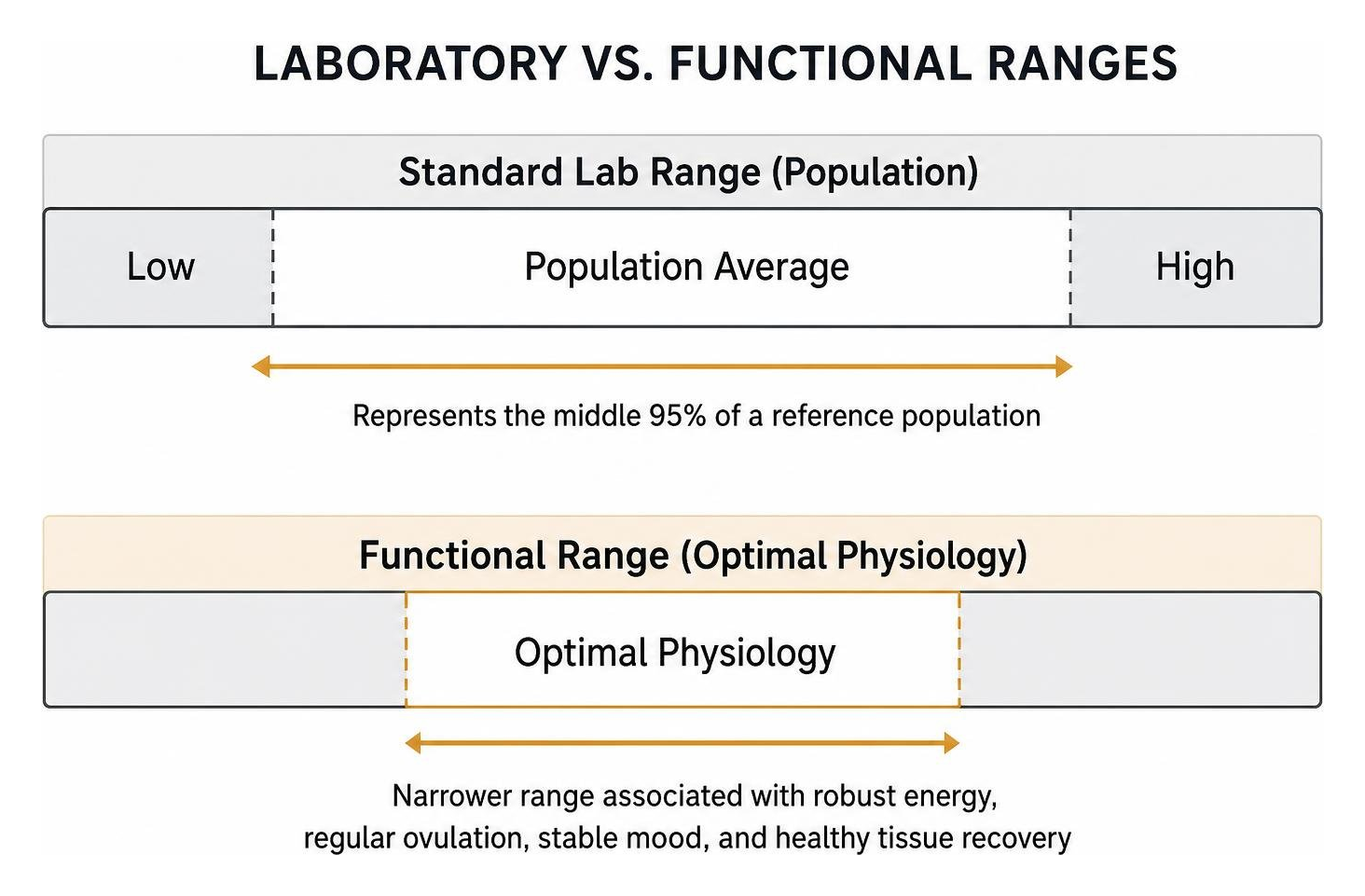
Reviewing a laboratory report can feel overwhelming. Understanding the distinction between standard population ranges and functional targets is key to making sense of your data.
Understanding Your Biomarker Data and Reference Ranges
Standard laboratory reference ranges represent a broad statistical average (the middle 95% of a reference population), which often includes individuals experiencing undiagnosed fatigue, metabolic strain, or subclinical hormone shifts. Optimal wellness ranges, by contrast, focus on the narrower bands associated with robust energy, regular ovulation, stable mood, and healthy tissue recovery.
When reviewing your markers, pay close attention to the following indicators:
Estradiol (E2): Baseline early follicular levels generally sit between 30 and 100 pg/mL. Mid-luteal levels often range from 100 to 250 pg/mL. Levels consistently below 30 pg/mL during reproductive years may indicate hypothalamic suppression or low energy availability.
Progesterone: A mid-luteal reading above 10 ng/mL strongly suggests successful ovulation. Levels below 5 ng/mL during the luteal phase point to anovulatory cycles or poor corpus luteum function.
FSH & LH: In the early follicular phase, balanced ratios sit close to 1:1 (typically 3 to 8 mIU/mL each). An LH-to-FSH ratio exceeding 2:1 or 3:1 can signal polycystic ovarian tendencies. FSH climbing above 15 to 20 mIU/mL on Day 3 warrants a closer look at ovarian reserve and the perimenopausal transition.
Total Testosterone: A typical healthy female range is roughly 15 to 70 ng/dL. Sensitive LC/MS-MS methods provide the most dependable readings at low concentrations.
Thyroid (TSH, Free T3, Free T4): While clinical TSH reference limits often extend up to 4.5 mIU/L, many individuals feel and perform best when TSH settles between 1.0 and 2.2 mIU/L, with Free T3 placed comfortably in the upper half of its range.
Connecting these insights to your everyday training and recovery is covered in our biomarker training plan guide 2026.
How to Interpret a Female Hormone Testing Panel with a Clinician
A single lab result is a snapshot of dynamic, living systems. When reviewing your panel with an experienced clinician:
Synthesize Labs with Symptoms: Review lab values alongside your cycle diary, basal body temperature trends, daily stress, and training logs.
Evaluate Biomarker Ratios: Look at relational metrics, such as the balance between mid-luteal estradiol and progesterone, or free testosterone relative to SHBG.
Address Foundational Drivers First: Nutrient insufficiencies (such as low vitamin D or ferritin), unmanaged systemic inflammation, and caloric deficits can suppress the neuroendocrine axis.
Establish a Structured Retesting Cadence: Schedule follow-up lab draws every 3 to 6 months to monitor how targeted nutritional, training, or clinical adjustments are working.
Frequently Asked Questions About Female Hormone Panels
How much does a comprehensive female hormone panel typically cost?
Direct-to-consumer and cash-pay clinical hormone panels generally range between $150 and $450, depending on marker depth. A basic 7-marker panel typically costs between $150 and $225, while expanded endocrine assessments that include complete thyroid panels and stress hormones run from $289 to $448. Most direct-access labs qualify for Health Savings Account (HSA) and Flexible Spending Account (FSA) reimbursement.
Can a hormone panel definitively diagnose menopause or PCOS?
A hormone panel cannot diagnose these conditions in isolation; it provides objective biomarker data that must be paired with clinical criteria. Under the Rotterdam criteria, diagnosing PCOS requires meeting at least two of three features: ovulatory dysfunction, clinical or biochemical signs of hyperandrogenism, and polycystic ovaries on ultrasound.
Similarly, menopause is defined clinically as 12 consecutive months without a menstrual period. While consistently elevated FSH levels (often >30 mIU/mL) alongside low estradiol support the diagnosis, hormone levels can fluctuate widely during perimenopause.
How does hormonal balance impact athletic recovery and body composition?
Hormones govern the balance between anabolic recovery and catabolic breakdown. Adequate estradiol and testosterone preserve muscle mass, support bone mineralization, and maintain tendon elasticity.
When energy availability drops—often seen in Relative Energy Deficiency in Sport (RED-s)—the body downregulates LH pulsatility, suppresses thyroid output, and elevates basal cortisol. This slows muscle protein synthesis, impairs glycogen replenishment, elevates injury risk, and halts training progression.
Conclusion
Taking a proactive approach to hormonal health helps you detect subtle physiological imbalances before they develop into chronic fatigue, cycle loss, or metabolic resistance. By tracking key reproductive, thyroid, and adrenal markers at the right moments in your cycle, you gain the clarity needed to personalize your training, nutrition, and daily recovery.
At Longevity Athlete, we believe objective data should always translate into clear, practical protocols. The Signature Plan is a self-guided group program. The Gold and Platinum Plans create individualized lifestyle protocols. When you combine systematic blood testing with expert interpretation, you can stop guessing and start building a balanced foundation for long-term health and performance.
To explore our full range of testing and optimization services, visit our home page.