Understanding What Your Inflammation Blood Markers Say About You
Core Inflammation Blood Markers and What They Measure
Inflammation blood markers measure the body's acute-phase response to cellular stress, trauma, or infection. Key markers like C-reactive protein (CRP), erythrocyte sedimentation rate (ESR), and GlycA indicate whether inflammation is acute (e.g., bacterial infection, severe injury) or chronic (e.g., low-grade vascular inflammation, cardiometabolic risk). Elevated CRP above 50 mg/L often signals severe bacterial infection or sepsis, while high-sensitivity CRP (hsCRP) between 1.0 and 3.0 mg/L reflects moderate cardiovascular risk. ESR rises more slowly and remains elevated for weeks, useful for monitoring autoimmune flares. GlycA provides a stable, multi-week average of systemic inflammation, resistant to short-term spikes. Understanding these patterns helps distinguish transient physiological responses from persistent, disease-driving inflammation.
When tissues experience cellular stress, trauma, or invasion by pathogens, immune cells dispatch pro-inflammatory signaling cytokines like interleukin-6 (IL-6), interleukin-1 beta (IL-1β), and tumor necrosis factor-alpha (TNF-α). These signaling molecules travel via the bloodstream directly to the liver hepatocytes, initiating the synthesis of acute-phase proteins (Singh et al., 2025). Understanding how these proteins behave is essential for tracking both acute physiological reactions and smoldering systemic issues (Amezcua-Castillo et al., 2023). You can review a comprehensive guide to C-reactive protein interpretation for an in-depth clinical framework.
Biomarker | Primary Clinical Indications | Kinetics (Rise / Peak / Clearance) | Standard Reference Range |
|---|---|---|---|
Standard CRP | Acute bacterial infection, severe trauma, post-surgical monitoring, sepsis | Rises: 6–12 hrs; Peak: 24–48 hrs; Halflife: ~19 hrs | Under 5.0 mg/L (or under 0.5 mg/dL) |
High-Sensitivity CRP (hsCRP) | Low-grade vascular inflammation, cardiovascular risk stratification | Continuous steady-state reflecting chronic vascular stress | Under 1.0 mg/L (Low Risk), 1.0–3.0 mg/L (Moderate), Above 3.0 mg/L (High) |
ESR (Sed Rate) | Autoimmune flare-ups (lupus, polymyalgia rheumatica), osteomyelitis | Rises: 24–48 hrs; Peak: Several days; Clearance: Weeks | Men: under 15–20 mm/hr; Women: under 20–30 mm/hr |
GlycA / GlycB | Chronic metabolic inflammation, cumulative cardiometabolic risk | Stable multi-week systemic integration; resistant to short-term spikes | Under 400 µmol/L (Optimal: under 300 µmol/L) |
Procalcitonin | Distinguishing severe bacterial infections from viral syndromes | Rises: 2–4 hrs; Peak: 6–24 hrs; Halflife: ~24 hrs | Under 0.1 µg/L (Normal/viral), Above 0.5 µg/L (Bacterial sepsis likely) |
C-Reactive Protein (CRP) and High-Sensitivity CRP (hsCRP)
In healthy individuals, C-reactive protein is merely a trace protein circulating at concentrations below 5 mg/L (or roughly 0.8–1.0 mg/dL). Because standard clinical laboratory assays historically struggled to measure tiny shifts at the bottom of that spectrum, high-sensitivity CRP (hsCRP) assays were developed to quantify low-grade inflammation down to fractional milligram levels.
For routine clinical evaluation, clinical testing guidelines for serum CRP help distinguish baseline fluctuations from severe pathology:
Healthy baseline: Under 1.0 mg/L (or under 0.1 mg/dL)
Minor/low-grade elevation: 1.0 to 3.0 mg/L (frequently linked to metabolic strain, smoking, mild physical trauma, or elevated cardiovascular risk)
Moderate systemic elevation: 10 to 50 mg/L (observed in active autoimmune conditions, myocardial infarction, or moderate tissue injury)
Severe acute elevation: Greater than 50 to 100+ mg/L (strongly associated with systemic bacterial infections in approximately 90% of clinical cases, or severe trauma and sepsis)
Pentameric vs. Monomeric CRP Isoforms
Under native physiological conditions, CRP circulates as a symmetrical, five-subunit ring known as pentameric CRP (pCRP) (Singh et al., 2025). In this baseline structural state, pCRP primarily performs homeostatic and host-defense functions, binding phosphocholine exposed on damaged cells to facilitate gentle, non-destructive clearance (Singh et al., 2025).
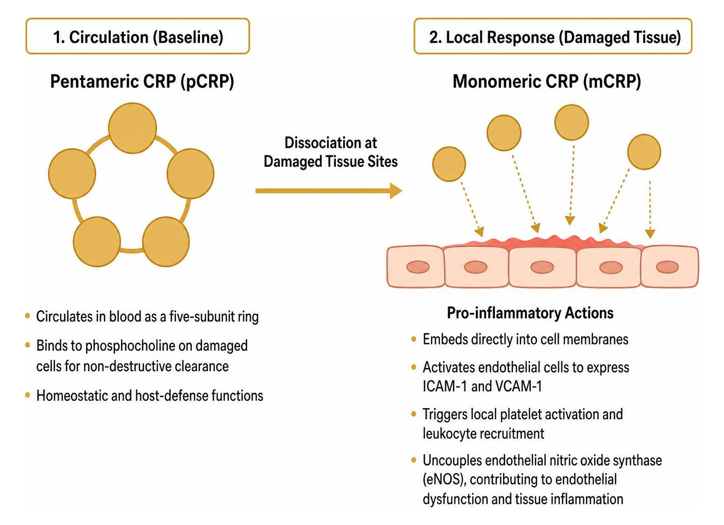
When pCRP encounters damaged cell membranes, activated platelets, or localized oxidative stress, it undergoes conformational dissociation into individual subunits called monomeric CRP (mCRP) (Singh et al., 2025). Unlike its circulating parent, mCRP is markedly pro-inflammatory:
It embeds directly into cell membranes.
It activates vascular endothelial cells to express adhesion molecules like ICAM-1 and VCAM-1.
It triggers local platelet activation and leukocyte recruitment.
It uncouples endothelial nitric oxide synthase (eNOS), directly contributing to endothelial dysfunction and localized tissue inflammation (Amezcua-Castillo et al., 2023).
Interpreting Novel NMR-Derived Inflammation Blood Markers
Nuclear Magnetic Resonance (NMR) spectroscopy now captures complex composite inflammatory signals that complement conventional single-protein tests. As detailed in a peer-reviewed study on NMR spectroscopy derived inflammatory biomarkers, these advanced markers evaluate systemic health through a broader biochemical lens:
GlycA & GlycB: These spectral peaks represent the aggregate signal of N-acetylglucosamine and N-acetylneuraminic acid residues on acute-phase circulating glycoproteins (including alpha-1-acid glycoprotein, haptoglobin, and alpha-1-antitrypsin). GlycA reflects cumulative, multi-pathway systemic inflammation and provides superior long-term stability compared to the acute fluctuations of standard CRP.
Supramolecular Phospholipid Composite (SPC): Derived from lipoprotein-associated phospholipid sub-regions, SPC signals capture lipid-driven inflammatory dynamics. Notably, SPC 2 (an HDL-related phospholipid marker) is significantly higher in women across all age brackets—averaging 33.7% higher than in men—reflecting estrogen-mediated effects on hepatic lipid processing.
Adiposity Dynamics: While GlycA and GlycB rise with each incremental increase in BMI class, SPC 2 displays a marked inverse correlation with BMI, dropping substantially in individuals with obesity.
Clinical Significance: What Elevated Markers Reveal About Your Health
Systemic inflammation is the unifying driver across modern chronic illnesses. An isolated spike in an acute-phase protein acts as an alert system, but persistent, unaddressed low-grade inflammation damages vascular beds, degrades musculoskeletal tissues, and accelerates cellular aging.
Cardiovascular Disease and Atherosclerosis
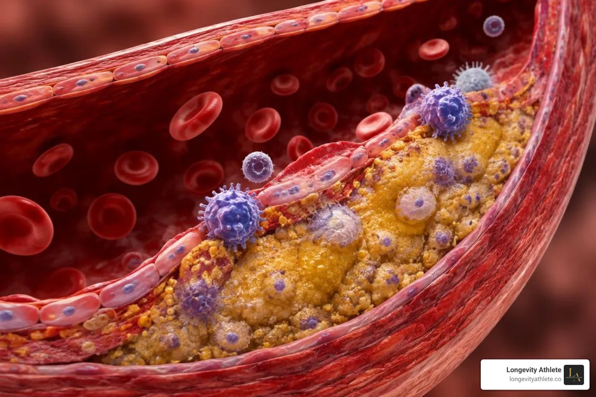
Cardiovascular disease is an active inflammatory process rather than a simple plumbing problem of passive cholesterol accumulation (Amezcua-Castillo et al., 2023). Higher circulating CRP levels act as independent risk factors for cardiac events and mortality (Amezcua-Castillo et al., 2023). Reviewing the evidence on CRP as a marker in coronary artery disease illustrates how localized vascular inflammation drives atheroma formation and destabilizes fibrous caps.
When assessing cardiovascular risk in asymptomatic individuals:
hsCRP under 1.0 mg/L: Low relative cardiovascular risk
hsCRP 1.0 to 3.0 mg/L: Moderate relative risk
hsCRP greater than 3.0 mg/L: High relative risk (conferring up to an 80% increase in relative coronary risk compared to baseline quintiles) (Amezcua-Castillo et al., 2023)
Cardiovascular risk scoring algorithms like the Reynolds Risk Score integrate hsCRP alongside total cholesterol, HDL-C, and blood pressure, successfully reclassifying clinical risk in up to 50% of intermediate-risk individuals (Amezcua-Castillo et al., 2023).
Autoimmune Disorders, Infections, and Chronic Disease States
The magnitude and kinetic profile of inflammatory markers provide direct clues about the underlying physiological challenge:
Bacterial vs. Viral Infections: Severe bacterial infections routinely trigger dramatic hepatic synthesis of CRP, driving serum concentrations above 50–100 mg/L within 24 to 48 hours (Singh et al., 2025). Viral infections, by contrast, stimulate interferon-alpha pathways that suppress hepatic CRP production, generally resulting in modest elevations (under 20 mg/L).
Autoimmune Activity: Conditions like rheumatoid arthritis, polymyalgia rheumatica, and inflammatory bowel disease exhibit sustained elevations in both CRP and ESR during active flares. Interestingly, in systemic lupus erythematosus (SLE), circulating CRP often remains disproportionately normal or only mildly elevated despite severe disease activity (unless serositis or concurrent infection is present).
Oncology and Frailty: Chronically elevated baseline hsCRP directly correlates with an increased rate of physical frailty progression in aging populations and is epidemiologically associated with higher risks of colorectal, gastric, and lung malignancies.
Non-Pathological Modifiers and Confounding Variables
Biomarker interpretation requires adjusting for personal demographics and baseline physiology:
Biological Sex and Hormones: Baseline hsCRP and NMR-derived SPC 2 markers are naturally higher in females due to endogenous estrogen activity and oral contraceptive or hormone replacement use.
Adiposity and BMI: Adipose tissue is an active endocrine organ. Adipocytes continuously secrete IL-6, elevating baseline CRP and GlycA in individuals with higher visceral body fat even in the absence of infection.
Age and Genetics: Baseline inflammatory markers drift upward with advancing age (termed inflammaging). Furthermore, common single nucleotide polymorphisms (SNPs) in the CRP gene alter baseline hepatic output independently of disease status (Singh et al., 2025).
Liver Function: Because the liver manufactures acute-phase reactants, advanced hepatic impairment or cirrhosis can blunt the synthesis of CRP, producing falsely reassuring, low values despite active systemic infection (Singh et al., 2025).
How Lifestyle and Medications Modulate Inflammation Blood Markers
Intense Exercise: Strenuous endurance exercise or heavy eccentric resistance training creates transient microtrauma in skeletal muscle, triggering a sharp IL-6 surge that causes CRP to spike within 12–24 hours post-workout before returning to baseline during recovery.
Sleep and Psychological Stress: Chronic sleep restriction (less than 6 hours per night) and severe emotional stress elevate nocturnal sympathetic tone and upregulate baseline hsCRP.
Dietary Factors: Diets rich in polyphenols, omega-3 fatty acids, and dietary fiber systematically lower circulating hsCRP and GlycA over multi-week timeframes.
Pharmacology: Common medications modify inflammatory readouts. Statins and GLP-1 receptor agonists exert powerful anti-inflammatory effects that significantly depress hsCRP independently of lipid changes (Amezcua-Castillo et al., 2023). Biologic agents (especially IL-6 receptor antagonists like tocilizumab) can completely suppress CRP synthesis (Singh et al., 2025).
Emerging Point-of-Care Assays and Testing Innovations
The timeline for inflammation testing has shifted rapidly from multi-day central laboratory turnarounds to near-instantaneous bedside precision:

Microfluidic CRP-Chips: Utilizing paper-based microfluidics and smartphone optical readers, modern decentralized testing devices quantify capillary-blood CRP in under five minutes.
Rapid Immunoassay Platforms: Systems like ProciseDx enable point-of-care, laboratory-grade quantification of acute-phase proteins directly in clinical or athletic training environments, streamlining real-time monitoring of recovery kinetics and infection control.
Integrating Biomarker Panels into Personalized Health Strategies
Tracking inflammation blood markers is most valuable when integrated into a structured, longitudinal health program. Relying on a single test in isolation leaves you vulnerable to misinterpreting transient post-exercise spikes or missing silent, low-grade metabolic distress.
To optimize your systemic health, we recommend the following testing best practices:
Establish True Baseline Measurements: Always collect routine inflammatory panels when well-rested, at least 48 hours away from exhausting athletic training, and free from acute viral or dental symptoms.
Combine Multi-Analyte Markers: Pair standard hsCRP with GlycA, full lipid profiles, fasting insulin, and hematological indices to distinguish isolated physical stress from systemic metabolic inflammation.
Track Kinetic Trends: If an unexpected elevation occurs, repeat the test in 2–3 weeks to establish whether the reading was an acute physiological response or a persistent elevation requiring targeted intervention.
Our expert biomarker testing services analyze complete physiological profiles to remove guesswork from health optimization. If you prefer a collective approach, you can join the self-guided group program in the Signature Plan. For high-touch support, our team builds individualized lifestyle protocols through the Gold Membership and delivers comprehensive executive oversight with individualized lifestyle protocols through the Platinum Membership.
Frequently Asked Questions About Blood Markers for Inflammation
How quickly do inflammation markers rise and normalize after injury or infection?
CRP concentrations begin to rise within 6 to 12 hours following an acute tissue injury or infectious stimulus, reaching peak circulating levels between 24 and 48 hours (Singh et al., 2025). Because circulating CRP has a plasma half-life of approximately 19 hours, levels decline quickly once the inciting inflammatory event is resolved (Singh et al., 2025). In contrast, markers like ESR can take several days to peak and multiple weeks to return to normal.
What is the difference between standard CRP and hsCRP tests?
Both tests measure the exact same protein produced by the liver. The difference lies entirely in assay sensitivity. A standard CRP test is designed to measure marked elevations ranging from 5 mg/L to over 100 mg/L, making it ideal for detecting acute bacterial infections, surgical complications, and autoimmune flares (Singh et al., 2025). An hsCRP test uses laser nephelometry or high-sensitivity immunoassays to accurately measure minute concentrations between 0.1 and 5.0 mg/L, which is necessary for evaluating low-grade vascular inflammation and cardiovascular risk (Amezcua-Castillo et al., 2023).
Can intense exercise cause temporary spikes in inflammatory markers?
Yes. Heavy eccentric resistance exercise, ultra-endurance running, or prolonged high-intensity training causes micro-damage in muscle fibers. This prompts local macrophages and myocytes to release interleukin-6, driving a temporary increase in hepatic CRP production over the subsequent 24 to 48 hours. When testing baseline inflammatory health, athletes should avoid strenuous workouts for 48 hours prior to phlebotomy.
Conclusion
Inflammation is not an enemy to be eliminated—it is an essential biological survival system that repairs damaged tissue and neutralizes pathogens (Singh et al., 2025). The problem arises when low-grade inflammation burns unchecked beneath the surface, degrading blood vessels, impairing physical recovery, and accelerating aging (Amezcua-Castillo et al., 2023).
At Longevity Athlete, we deliver the precision biomarker tracking, advanced data interpretation, and clinical expertise needed to help you take full control of your health. Whether you choose our self-guided group programming or our high-touch executive coaching, our protocols turn complex laboratory data into clear, actionable strategies. Ready to optimize your biological longevity? Visit our site to explore tailored performance and longevity protocols today.
References
Baltej Singh, Amandeep Goyal, Bhupendra C. Patel. "C-Reactive Protein: Clinical Relevance and Interpretation." StatPearls Publishing, 2025. NBK441843. Amezcua-Castillo E, González-Pacheco H, Sáenz-San Martín A, Méndez-Ocampo P, Gutierrez-Moctezuma I, Massó F, Sierra-Lara D, Springall R, Rodríguez E, Arias-Mendoza A, Amezcua-Guerra LM. "C-Reactive Protein: The Quintessential Marker of Systemic Inflammation in Coronary Artery Disease-Advancing toward Precision Medicine.." Biomedicines, 2023. PMCID PMC10525787.