Inflammation Markers 101
Understanding Key Inflammation Markers in the Human Body
When cells experience trauma, oxidative stress, or invasion by pathogens, your immune system launches an inflammatory cascade. Immune cells release signaling cytokines—primarily interleukin-6 (IL-6), interleukin-1 beta (IL-1β), and tumor necrosis factor-alpha (TNF-α). These cytokines travel through the bloodstream to the liver, where they trigger the rapid production and release of acute-phase reactants (NBK519570).
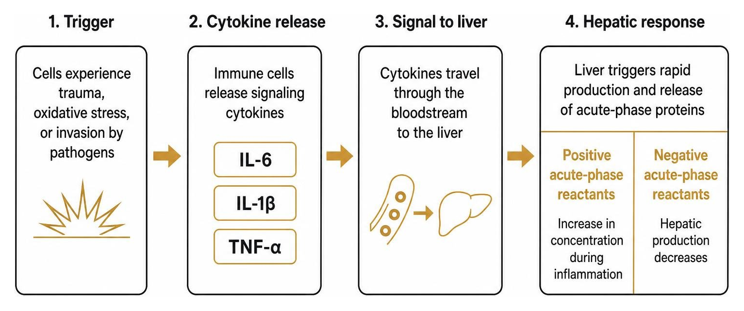
These circulating proteins fall into two categories:
Positive acute-phase reactants: Serum proteins that increase in concentration during inflammation (such as C-reactive protein, ferritin, fibrinogen, procalcitonin, and hepcidin).
Negative acute-phase reactants: Proteins whose hepatic production decreases as the liver prioritizes defense molecules (notably albumin and transferrin).
Clinicians and performance specialists track these proteins to gauge the intensity and resolution of immune activity, as outlined in clinical research on acute phase reactants.
Marker | Biological Source | Primary Kinetic Profile | Primary Clinical Utility |
|---|---|---|---|
C-Reactive Protein (CRP) | Hepatic synthesis via IL-6 | Rises in 4–6 hrs; peaks at 36–50 hrs; 19-hr half-life | General systemic inflammation, infection, cardiovascular risk |
Erythrocyte Sedimentation Rate (ESR) | Indirect (erythrocyte aggregation via fibrinogen) | Rises in 24–48 hrs; slow multi-day resolution | Chronic inflammatory conditions, temporal arteritis, polymyalgia |
Procalcitonin (PCT) | Thyroid C-cells / parenchymal tissue via bacterial endotoxins | Rises in 2–4 hrs; peaks at 12–24 hrs | Distinguishing severe bacterial infections from viral or sterile causes |
Ferritin | Reticuloendothelial system / hepatocytes | Moderate rise; prolonged plateau | Iron storage assessment, macrophage activation, chronic disease monitoring |
C-Reactive Protein (CRP) vs. Erythrocyte Sedimentation Rate (ESR)
While clinicians often order CRP and ESR together, they evaluate biological activity through different mechanisms.
CRP is a direct, positive acute-phase protein synthesized by the liver. Its plasma concentrations rise within 4 to 6 hours after an acute stimulus, doubling roughly every 8 hours before peaking around 36 to 50 hours. Because CRP possesses a constant plasma half-life of 19 hours, its concentration drops quickly once tissue damage or infection resolves.
By contrast, ESR is an indirect measurement of inflammation. It measures the distance (in millimeters) that red blood cells settle in an anticoagulated tube of blood over one hour. During systemic inflammation, elevated levels of positively charged plasma proteins (primarily fibrinogen) shield the negative zeta potential of red blood cells. This neutralizes their natural repulsion, allowing them to stack into coin-like structures called rouleaux that fall rapidly to the bottom of the tube.
Because fibrinogen takes 24 to 48 hours to rise and clears much slower than CRP, ESR exhibits longer diagnostic latency. Evaluating standard inflammation blood markers requires recognizing that ESR can remain elevated for days or weeks after an acute illness has resolved, whereas CRP directly mirrors current inflammatory status.
Procalcitonin, Ferritin, and Cytokines as Inflammation Markers
Evaluating complex health challenges often requires looking beyond CRP and ESR to specialized markers:
Procalcitonin (PCT): Under normal conditions, thyroid C-cells process procalcitonin into calcitonin. During systemic bacterial infections, bacterial endotoxins and pro-inflammatory cytokines trigger extrathyroidal tissues throughout the body to release intact procalcitonin directly into the blood. Because viral infections trigger interferon-gamma (IFN-γ)—which actively suppresses procalcitonin synthesis—PCT concentrations above 0.25 to 0.50 µg/L provide strong diagnostic evidence of bacterial rather than viral etiology.
Ferritin: While best known as an intracellular iron-storage molecule, ferritin also acts as a positive acute-phase reactant. During inflammatory states, hepatocytes and macrophages upregulate ferritin synthesis to sequester free serum iron, depriving invading pathogens of an essential nutrient.
Direct Cytokines (IL-6, TNF-α, IL-1β): Upstream signaling molecules like IL-6 can be measured directly, though their short circulatory half-lives make downstream hepatic proteins like CRP more stable for regular monitoring.
C-Reactive Protein Deep Dive: Biology, Isoforms, and Reference Ranges
C-reactive protein was discovered in 1930 when researchers identified a serum substance that precipitated the C-polysaccharide component of the Streptococcus pneumoniae cell wall. Far from being a simple, inert marker, CRP plays an active role in innate immunity by binding phosphocholine residues exposed on damaged host cell membranes and foreign pathogens (NBK441843).
At the molecular level, CRP exists in two biologically distinct structural isoforms, as detailed in the StatPearls guide to C-reactive protein:
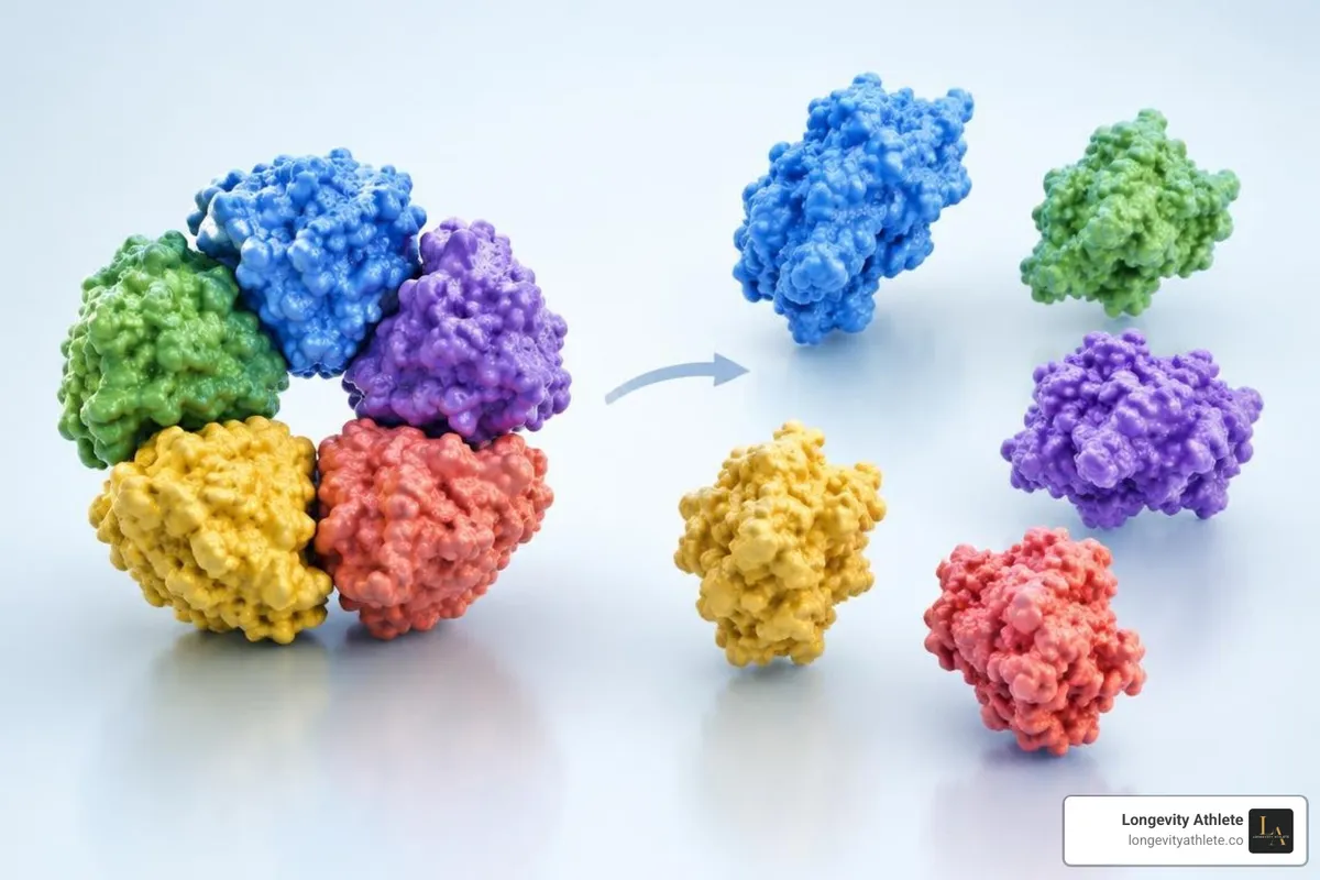
Pentameric CRP (pCRP): The native, disk-shaped molecule that circulates in healthy plasma. It consists of five identical, non-covalently linked 23-kDa globular subunits arranged symmetrically around a central pore (total molecular weight ~115 kDa). Native pCRP exerts net anti-inflammatory properties, maintaining vascular homeostasis and dampening excessive complement activation.
Monomeric CRP (mCRP): When circulating pCRP binds to damaged cell membranes, activated platelets, or apoptotic cellular debris in the presence of calcium and localized acidic pH, its pentameric ring dissociates into individual, pro-inflammatory monomeric subunits. These mCRP subunits promote leukocyte recruitment, trigger platelet aggregation, stimulate endothelial dysfunction, and accelerate reactive oxygen species generation.
Standard CRP vs. High-Sensitivity CRP (hs-CRP)
The standard CRP test and the high-sensitivity CRP (hs-CRP) test measure the same protein, but their analytical detection limits differ significantly:
Standard (Conventional) CRP: Uses traditional turbidimetric or nephelometric assays designed to detect gross elevations, with an analytical range between 3.0 and 1,000 mg/L (0.3 to 100 mg/dL). It is primarily used to identify acute bacterial infections, systemic autoimmune flares, major tissue trauma, or post-surgical healing.
High-Sensitivity CRP (hs-CRP): Utilizes laser nephelometry, high-precision immunoturbidimetry, or enzyme-linked immunosorbent assays (ELISA) to detect minute concentrations between 0.1 and 10.0 mg/L. This lower threshold allows clinicians to detect low-grade, subclinical vascular inflammation that would register as an undetectable baseline reading on a standard assay.
For disciplined individuals tracking long-term metabolic health, comprehensive inflammation testing using hs-CRP reveals subtle changes that standard clinical panels overlook.
Interpreting Normal and Elevated Blood Ranges
In healthy young adults, the median baseline CRP concentration is approximately 0.8 mg/L, with the 90th percentile reaching 3.0 mg/L and the 99th percentile reaching 10.0 mg/L. Following a severe acute-phase trigger, circulating CRP levels can surge 10,000-fold from under 50 μg/L to more than 500 mg/L.
Low Cardiovascular Risk (hs-CRP): < 1.0 mg/L
Moderate Cardiovascular Risk (hs-CRP): 1.0 to 3.0 mg/L
High Cardiovascular Risk (hs-CRP): > 3.0 mg/L
Minor / Low-Grade Systemic Elevation: 3.0 to 10.0 mg/L (0.3 to 1.0 mg/dL) — Frequently linked to metabolic syndrome, central adiposity, subclinical gingivitis, smoking, or chronic lifestyle stressors.
Moderate Systemic Elevation: 10.0 to 100.0 mg/L (1.0 to 10.0 mg/dL) — Commonly observed in active autoimmune diseases (e.g., rheumatoid arthritis), myocardial infarction, deep vein thrombosis, and mild-to-moderate viral or localized bacterial infections.
Marked / Severe Elevation: > 100.0 mg/L (> 10.0 mg/dL) — Strongly associated with systemic bacterial infections (sepsis), major physical trauma, severe burns, or widespread necrotizing vasculitis. When concentrations exceed 500 mg/L (50 mg/dL), acute bacterial etiology accounts for approximately 90% of cases.
Clinical Applications: Cardiovascular Risk, Autoimmunity, and Disease
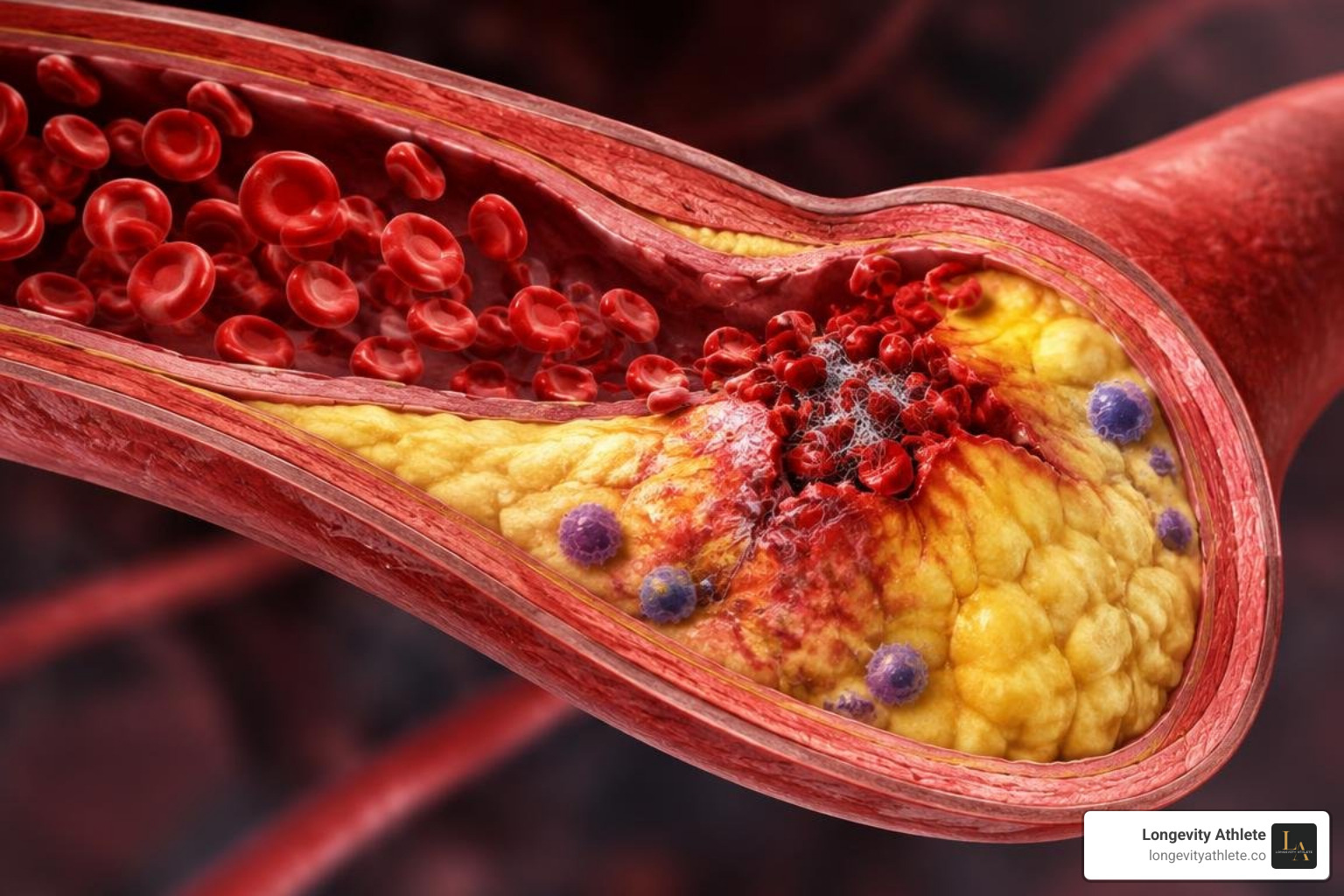
Elevated inflammation levels are closely linked with the initiation, progression, and rupture of atherosclerotic plaques. Circulating mCRP localizes directly within vascular lesions, impairing endothelial nitric oxide synthase (eNOS) transcription and upregulating adhesion molecules that pull monocytes into the arterial subendothelium.
Beyond coronary artery disease, elevated baseline CRP drives the pathogenesis of several chronic conditions:
Venous Thromboembolism (VTE): Elevated CRP promotes a prothrombotic state by upregulating tissue factor expression and downregulating endothelial protein C pathways.
Heart Failure: Chronic low-grade inflammation drives myocardial remodeling, progressive fibrosis, and diastolic stiffness.
Frailty Syndrome: Age-associated inflammatory signaling accelerates sarcopenia, impairs mitochondrial biogenesis in skeletal muscle, and exacerbates neurocognitive decline.
Diagnostic Role of Inflammation Markers in Autoimmune and Cardiovascular Disease
In clinical rheumatology, CRP provides a sensitive way to track disease activity and therapeutic response in conditions like rheumatoid arthritis (RA), where joint synovitis correlates with acute-phase protein output.
Interestingly, patients with active systemic lupus erythematosus (SLE) frequently present with disproportionately modest CRP elevations despite severe clinical flares, unless accompanied by concurrent bacterial infection or serositis. This occurs partly because high levels of interferon-alpha in SLE actively suppress hepatic CRP transcription.
In cardiology, hs-CRP serves as an independent prognostic marker for future cardiovascular events. Following an acute myocardial infarction (AMI), CRP levels appear within 1 to 2 days, peak precisely at day 3, and return toward baseline after day 7 as myocardial tissue repair stabilizes. Tracking these patterns helps clinicians evaluate the anti-inflammatory efficacy of lipid-lowering therapies like statins.
Differentiating Infectious, Inflammatory, and Tissue Trauma Triggers
Because acute-phase proteins react non-specifically to various biological stressors, evaluating the complete clinical context is essential:
Clinicians also rely on calculated biomarker ratios. For example, the CRP-to-albumin ratio predicts acute pancreatitis severity with high accuracy. Additionally, in cases of acute gallbladder inflammation, a CRP level of 110.5 mg/L or higher serves as an independent predictor of hospital stays lasting 7 days or longer.
Testing Modalities, Athletic Recovery, and Influencing Factors
Standard venous phlebotomy using automated laser nephelometry in reference laboratories remains the gold standard for clinical measurement. However, point-of-care testing (POCT) methods—including capillary fingerstick cassettes and microfluidic chips—can deliver reliable results in under five minutes. While POCT devices match laboratory accuracy for identifying gross bacterial thresholds (>20–50 mg/L), central laboratory nephelometry remains the preferred method for detecting subtle changes in high-sensitivity cardiovascular ranges (<3.0 mg/L).
For endurance athletes and strength practitioners, training volume directly impacts circulating performance biomarkers. Strenuous, unaccustomed eccentric exercise triggers transient structural microtrauma, elevating systemic inflammation markers alongside muscle enzyme markers like those seen on a creatine kinase blood test. When interpreting inflammatory biomarker results, blood draws must be scheduled during metabolically stable recovery windows to avoid misinterpreting exercise-induced recovery spikes as systemic pathology.
Modulating Factors: Medications, Diet, and Genetics
Interpreting systemic biomarkers accurately requires accounting for external and genetic variables:
Pharmaceutical Suppression: Nonsteroidal anti-inflammatory drugs (NSAIDs), systemic corticosteroids, HMG-CoA reductase inhibitors (statins), and targeted biologic therapies (such as IL-6 receptor antagonists like tocilizumab) significantly suppress CRP production.
Dietary Influences: Pro-inflammatory diets high in industrial trans-fatty acids and refined carbohydrates elevate circulating baselines. In a study of over 700 nurses, those in the highest quartile of trans fat consumption exhibited blood levels of CRP that were 73% higher than those in the lowest quartile. Dietary changes should be discussed with a qualified clinician or registered dietitian when inflammation or cardiometabolic risk is a concern.
Adiposity and Sleep: Adipose tissue—especially visceral fat—secretes steady amounts of IL-6, causing baseline CRP elevations (typically 1.0 to 4.0 mg/L) in individuals with obesity. Chronic sleep deprivation, obstructive sleep apnea, and psychosocial stress independently elevate circulating markers.
Genetics: Single nucleotide polymorphisms (SNPs) within the CRP gene on chromosome 1 (1q23.2) can alter baseline protein expression, shifting an individual's personal reference point.
Patient Education and Preparation Considerations
To ensure testing accuracy, patients and athletes should follow these guidelines:
Ensure Metabolic Stability: Postpone cardiovascular hs-CRP testing for at least two to three weeks following an acute viral infection, minor surgical procedure, or dental intervention.
Standardize Phlebotomy Protocol: Schedule blood draws in the morning during a well-hydrated, rested state. Avoid strenuous training sessions for 48 hours prior to testing.
Confirm with Serial Retesting: Because inflammatory levels fluctuate, isolated hs-CRP elevations in the 3.0 to 10.0 mg/L range should be re-tested after two to four weeks before adjusting clinical or training protocols. People using at-home testing should follow the collection instructions provided by the testing service.
Frequently Asked Questions About Inflammation Testing
What is the primary difference between standard CRP and hs-CRP tests?
Standard CRP and hs-CRP tests quantify the exact same hepatic protein, but they utilize assays with different analytical sensitivity ranges. Standard CRP tests detect gross elevations between 3.0 and 1,000 mg/L to diagnose acute bacterial infections, autoimmune conditions, or trauma. High-sensitivity CRP (hs-CRP) tests measure fine-grain concentrations between 0.1 and 10.0 mg/L, making them suitable for evaluating low-grade vascular inflammation and cardiovascular risk.
How quickly do CRP levels normalize after acute inflammation resolves?
Because circulating CRP possesses a constant plasma half-life of 19 hours across all medical conditions, serum levels decline rapidly once the underlying inflammatory trigger is cleared. Following uncomplicated infections or minor surgical procedures, concentrations generally return to baseline within 7 to 10 days.
Can strenuous exercise temporarily elevate systemic inflammation markers?
Yes. Intense, unaccustomed eccentric exercise or prolonged endurance events induce transient skeletal muscle microtrauma. This prompts localized IL-6 release and drives a temporary acute-phase reaction, causing CRP levels to peak 24 to 48 hours post-exercise before resolving.
Conclusion
Understanding systemic biomarker tracking allows athletes and health-conscious individuals to move beyond guesswork. By measuring foundational molecules like C-reactive protein, erythrocyte sedimentation rate, and procalcitonin in their proper clinical context, you gain clear visibility into tissue recovery, metabolic balance, and cardiovascular health.
At Longevity Athlete, we provide precision-performance wellness consulting for disciplined athletes and individuals who refuse to settle for generic health advice. The Signature Plan is a self-guided group program. The Gold and Platinum Plans create individualized lifestyle protocols built around comprehensive biomarker analysis and ongoing data-driven adjustments.
Visit Longevity Athlete to explore our tailored biomarker programs and take a data-driven approach to your health, recovery, and long-term performance.