
The Complete Guide to Inflammation Testing Options
Core Biomarkers Used in Clinical Inflammation Testing
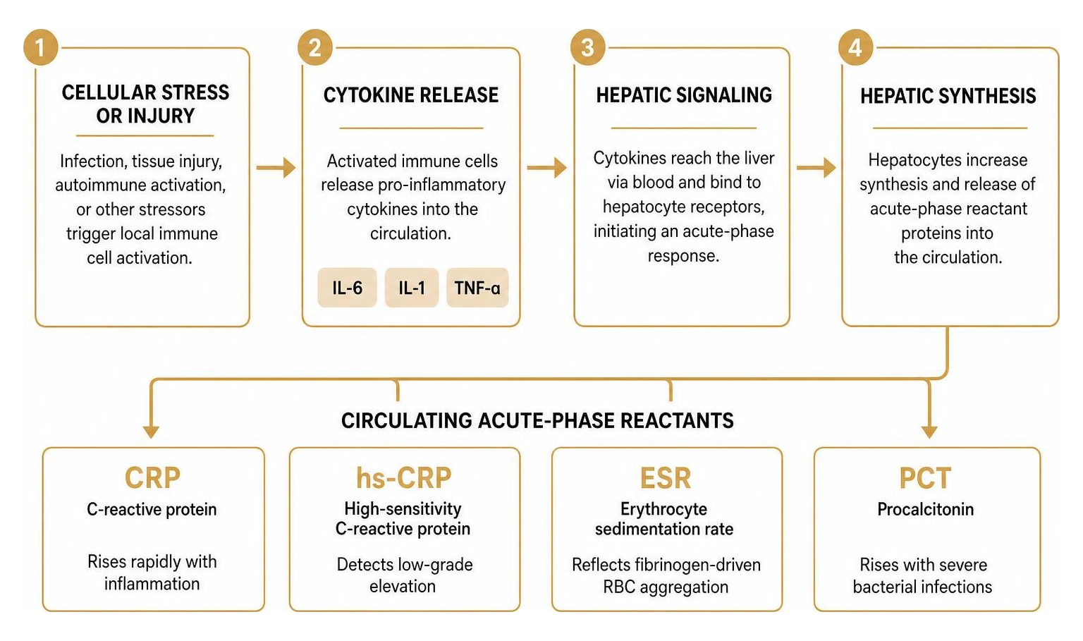
When clinicians evaluate systemic inflammation, they turn to circulating blood proteins known as acute-phase reactants. These proteins are produced primarily by the liver in direct response to inflammatory cytokines like interleukin-6 (IL-6), interleukin-1 (IL-1), and tumor necrosis factor-alpha (TNF-α).
Understanding the core differences between standard diagnostic markers ensures we choose the right tool for the right clinical question.
Biomarker | Diagnostic Utility | Primary Stimulus | Half-Life | Key Limitations |
|---|---|---|---|---|
Standard CRP | Acute tissue injury, infections, autoimmune flares | Hepatic synthesis via IL-6 | ~19 hours | Non-specific; spikes from minor trauma or intense training |
hs-CRP | Micro-vascular inflammation, cardiovascular risk stratification | Low-grade basal vascular inflammation | ~19 hours | Easily skewed by acute illness, minor injuries, or high-volume workouts |
ESR | Subacute and chronic inflammatory disease, monitoring rheumatologic conditions | Fibrinogen-driven red blood cell aggregation | Days to weeks | Confounded by age, female sex, anemia, obesity, and ambient temperature |
Procalcitonin (PCT) | Differentiating severe bacterial infections from viral illness | Extrathyroidal neuroendocrine tissue activation via endotoxins | ~24 hours | Low utility for localized, mild, or non-bacterial chronic inflammatory states |
Standard laboratory inflammation monitoring tests measure acute-phase reactants to capture active immune responses. C-reactive protein (CRP) is a pentameric protein that can rise more than 1,000-fold within 24 to 48 hours of an acute insult.
High-sensitivity CRP (hs-CRP) uses laser nephelometry or turbidimetry to detect tiny amounts of CRP below standard diagnostic thresholds. While routine CRP helps detect infections and major tissue damage, hs-CRP stratifies baseline cardiovascular risk into clear categories:
Low Risk: < 1.0 mg/L
Average Risk: 1.0 to 3.0 mg/L
High Risk: > 3.0 mg/L
Erythrocyte Sedimentation Rate Versus CRP Dynamics
Erythrocyte sedimentation rate (ESR) measures the distance in millimeters that red blood cells settle in an anticoagulated tube of whole blood over one hour. The Westergren method remains the classic standard, though modern clinical laboratories also use automated EDTA-based closed systems.
Under normal circumstances, red blood cells carry a net negative surface charge (zeta potential) that keeps them repelling one another. When inflammation introduces large, positively charged plasma proteins—such as fibrinogen and immunoglobulins—these charges are neutralized. The red blood cells stick together in stacks like dropped coins (rouleaux formation), increasing their mass and settling faster through the plasma.
However, ESR comes with notable technical and biological limitations:
Confounding Factors: ESR is heavily distorted by red blood cell morphology, ambient room temperature, hemoglobin levels (anemia artificially accelerates ESR), renal dysfunction, heart failure, and elevated BMI. In fact, labs must carefully regulate room temperatures because warmer environments artificially increase sedimentation rates.
Slow Clearance: While CRP normalizes rapidly once an inflammatory trigger resolves, fibrinogen clears slowly. This leaves ESR elevated for weeks after clinical recovery.
Diagnostic Precision: In a massive cohort study of 136,961 patients undergoing inflammatory testing, CRP demonstrated superior diagnostic performance with an Area Under the Curve (AUC) of 0.682, compared to an AUC of just 0.589 for ESR. Adding ESR alongside CRP yielded a negligible AUC increase to 0.688, highlighting that routine concurrent testing rarely adds meaningful diagnostic value.
Procalcitonin and Acute Infection Markers
Procalcitonin (PCT) is the peptide precursor of calcitonin. Under baseline conditions, it is produced by thyroid C-cells and stays nearly undetectable in circulation.
During systemic bacterial infections, bacterial endotoxins and inflammatory cytokines trigger the CALC-1 gene across broad extrathyroidal tissues, causing a rapid surge of procalcitonin into the bloodstream within 2 to 4 hours.
Because viral infections stimulate interferon-gamma (IFN-γ)—which actively suppresses procalcitonin synthesis—PCT provides exceptional specificity in distinguishing bacterial infections from viral illnesses. Point-of-care infection and inflammation diagnostics measuring PCT help clinicians triage acute respiratory infections, manage sepsis protocols, identify early-onset neonatal sepsis, and support responsible antibiotic stewardship.
Clinical Indications and Diagnostic Guidelines for Inflammatory Markers

Clinical practice guidelines and insurer medical necessity frameworks outline clear rules for ordering CRP and ESR. Major health plans, such as Geisinger Health Plan, explicitly restrict reimbursement when CRP and ESR are ordered together for general symptoms, designating CRP as the preferred test due to its superior specificity, reproducibility, and lack of age- or sex-related skewing.
Rheumatoid Arthritis (RA): The American College of Rheumatology (ACR) and European Alliance of Associations for Rheumatology (EULAR) recommend baseline testing of CRP and ESR to calculate composite disease activity scores (such as DAS28-CRP). During active disease, monitoring is recommended every 1 to 3 months; once clinical remission or low disease activity is achieved, testing frequency shifts to every 6 to 12 months.
Hodgkin Lymphoma: The National Comprehensive Cancer Network (NCCN) includes an elevated ESR (≥50 mm/hr without B symptoms, or ≥30 mm/hr with B symptoms) as a key prognostic factor that designates early-stage disease as unfavorable, directly influencing chemotherapy and radiation protocols.
Autoimmune Conditions, Giant Cell Arteritis, and Polymyalgia Rheumatica
Giant cell arteritis (GCA) and polymyalgia rheumatica (PMR) are distinct exceptions where dual testing remains clinically justified:
In GCA, ESR alone is elevated in up to 89% of patients.
However, when both ESR and CRP are ordered together, sensitivity rises to 99% and specificity reaches 97%.
In pediatric populations, systemic autoimmune and inflammatory diseases present unique testing dynamics. A clinical study of 150 pediatric patients with systemic inflammatory diseases revealed that 73.3% presented with abnormal CRP, while 100% displayed abnormal ESR levels upon hospital admission. This demonstrates that prolonged immune activity in younger cohorts can drive broad plasma protein changes that make ESR a reliable screening partner.
Differentiating Septic Joints and Periprosthetic Joint Infections
In orthopedics, differentiating septic arthritis or periprosthetic joint infection (PJI) from aseptic joint failure is critical. In a meta-analysis of 30 studies covering 3,909 revision total hip and knee arthroplasties:
CRP demonstrated a diagnostic odds ratio of 13.1 for detecting periprosthetic joint infection.
ESR demonstrated a diagnostic odds ratio of 7.2.
A two-year retrospective clinical study of 163 patients presenting with acute joint effusion showed that mean CRP was 13 mg/dL in septic joints versus 8.5 mg/dL in non-septic joints, establishing CRP as a dependable marker for diagnosing acute septic joints. By contrast, mean ESR showed substantial overlap (57 mm/hr in septic joints versus 46 mm/hr in non-septic joints), offering limited clinical utility for acute differentiation.
Advanced Multiplex Panels and Emerging Biomarkers
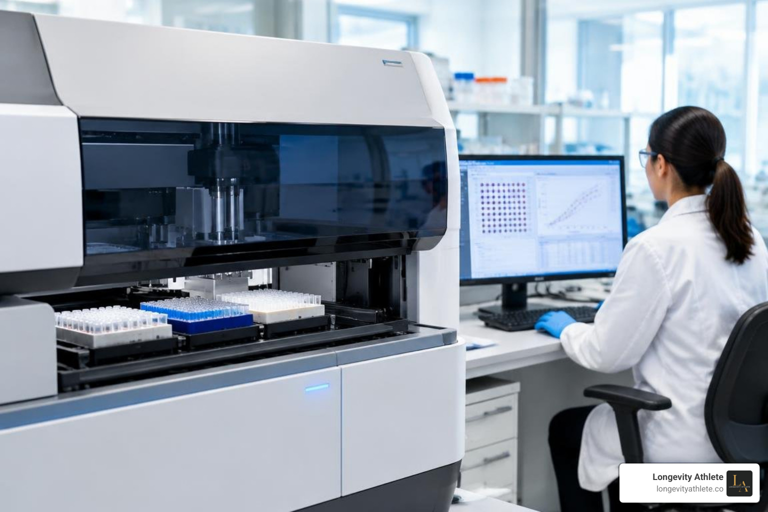
When routine tests leave clinical questions unanswered, advanced multiplex testing provides deeper insight into immune activity. Rather than measuring a single downstream acute-phase reactant, multiplex assays quantify dozens of upstream signaling molecules simultaneously.
For individuals seeking comprehensive oversight, our precision health services use targeted biomarker tracking to tailor training, nutrition, and recovery strategies to individual physiology.
Multiplex Assays and Cytokine Panels in Modern Inflammation Testing
Modern platforms utilize magnetic bead-based multiplex technology to measure up to 37 biomarkers in under four hours from a single micro-volume blood sample. Analyzed on specialized fluorescent suspension systems, these multiplex panels capture:
TNF Superfamily Proteins: sCD30, sCD163, APRIL, and BAFF, which reflect macrophage activation and B-cell signaling.
Interferon Family: IFN-α2, IFN-β, and IFN-γ, which regulate antiviral and cell-mediated pathways.
Treg Cytokines & Matrix Metalloproteinases (MMPs): Interleukins regulating immune tolerance alongside MMP-1, MMP-2, and MMP-3, which track extracellular matrix degradation and tissue remodeling.
Bone Turnover Modulators: Osteocalcin and osteopontin, connecting chronic low-grade inflammation to bone metabolic shifts.
Cellular Reactivity and Food Sensitivity Testing
Chronic systemic inflammation can also stem from innate immune activation triggered by non-IgE-mediated dietary antigens. Unlike standard antibody testing, cellular leukocyte reactivity assessments measure ex vivo white blood cell volume and size shifts after exposure to food and chemical antigens.
Because neutrophils and monocytes alter their physical dimensions upon degranulation and activation, cellular testing helps clinicians design targeted elimination and reintroduction protocols for complex inflammatory symptoms.
Novel Biomarkers: Interleukin-6, Galectin-3, and Pentraxin-3
Comprehensive testing panels incorporate both classical and emerging biomarkers to capture acute immune responses and chronic tissue changes:
Interleukin-6 (IL-6): The primary driver of hepatic acute-phase protein synthesis. Measuring IL-6 directly identifies cytokine signaling before downstream proteins spike.
Galectin-3: A beta-galactoside-binding lectin that drives macrophage migration, tissue remodeling, and systemic fibrosis, serving as an early indicator of cardiovascular and renal strain.
Pentraxin-3 (PTX3): A structural relative of CRP produced directly at the site of vascular inflammation by endothelial cells and macrophages, providing direct insight into local vascular stress.
Fibrinogen and Antinuclear Antibodies (ANA): Assessing coagulation factors alongside autoantibodies helps clinicians rule out underlying autoimmune activity and manage long-term vascular health.
Actionable Protocols: Integrating Inflammation Testing into Longevity Care
For athletes and active individuals, random biomarker testing provides little actionable value without an organized testing framework. Exploring our performance and recovery research and reading our evidence-based health articles highlights how strategic data tracking prevents chronic overreaching and promotes sustained performance.
Establish a True Resting Baseline: Draw blood when fully rested, at least 48 to 72 hours after strenuous exercise, free from acute illness or injury.
Track Trends Over Time: Monitor hs-CRP and upstream cytokines quarterly to evaluate whether changes in training volume, sleep quality, and diet are improving systemic recovery.
Differentiate Acute Muscle Damage from Chronic Strain: Creatine kinase (CK) spikes rapidly from local muscle fiber breakdown, whereas persistent elevations in hs-CRP and IL-6 reflect systemic recovery deficits.
Adjust Training and Recovery: When systemic inflammatory markers rise above baseline, reduce high-intensity training, prioritize sleep, and support recovery nutrition before structural overtraining occurs.
Frequently Asked Questions About Inflammation Testing Options
Why is CRP generally preferred over ESR for routine inflammation testing?
C-reactive protein is synthesized directly by hepatocytes in response to IL-6 and has a stable half-life of roughly 19 hours. It rises within hours of an inflammatory stimulus and falls rapidly as tissue heals.
By contrast, ESR relies on red blood cell aggregation driven by circulating fibrinogen. ESR is easily distorted by red blood cell shape, anemia, female sex, elevated BMI, and ambient lab temperatures, making CRP the more reliable and specific marker.
When should CRP and ESR be ordered together instead of individually?
Dual testing is clinically supported in conditions where combined sensitivity is needed. In suspected giant cell arteritis, ordering both tests increases diagnostic sensitivity to 99%.
Dual testing is also standard in orthopedic evaluations for periprosthetic joint infection and in pediatric rheumatology workups, where broad plasma protein shifts provide useful diagnostic context.
How often should inflammatory biomarkers be tested for health and performance tracking?
Active Autoimmune Disease: Every 1 to 3 months during active flares, tapering to every 6 to 12 months once remission is established.
Athletic Training: Quarterly testing during hard training blocks to monitor recovery, paired with an off-season baseline.
General Longevity & Wellness: Annual or semi-annual hs-CRP testing to assess baseline vascular health.
Conclusion
Systemic inflammation influences nearly every aspect of long-term health, cardiovascular resilience, and athletic performance. By moving past isolated blood draws and adopting proactive, trend-based biomarker tracking, you can pinpoint the underlying drivers of fatigue and optimize your recovery.
Ready to gain clearer visibility into your biomarkers? You can schedule a consultation with our team or contact our team directly to discuss our comprehensive longevity programs.
At Longevity Athlete, we specialize in personalized biomarker analysis and data-driven performance guidance. The Signature Plan is a self-guided group program. The Gold and Platinum Plans create individualized lifestyle protocols. Take control of your recovery and build lasting performance today.