Stop Guessing Your Inflammation Levels by Checking Key Blood Markers
Understanding the Primary Inflammatory Marker Blood Test Options: CRP vs. ESR
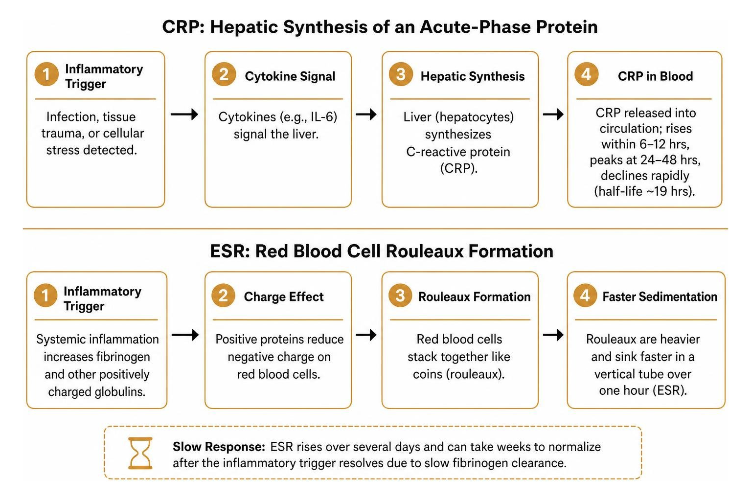
When your body detects cellular stress, an infection, or tissue trauma, the immune system initiates a complex signaling cascade. Circulating chemical messengers—primarily cytokines like interleukin-6 (IL-6)—signal the liver to synthesize acute-phase proteins. An inflammatory marker blood test captures these dynamic immune responses, helping clinicians distinguish between acute immune spikes and lingering, low-grade systemic stress.
The two main laboratory tools for assessing systemic inflammation are C-reactive protein (CRP) and the erythrocyte sedimentation rate (ESR). While both serve as indicators of immune activation, they measure different biological phenomena on completely different timelines.
CRP is an acute-phase protein synthesized directly by hepatocytes (liver cells). In response to systemic inflammatory triggers, CRP levels surge within 6 to 12 hours, reach peak concentrations around 24 to 48 hours, and decline rapidly once the trigger resolves due to its short plasma half-life (roughly 19 hours). You can review a detailed clinical overview of C-reactive protein testing to explore the biological mechanics of hepatic synthesis.
ESR, by contrast, is an indirect measurement of circulating plasma proteins—particularly fibrinogen. When systemic inflammation occurs, elevated levels of fibrinogen and other positively charged globulins cause negative charges on red blood cells (erythrocytes) to diminish. This allows the red cells to stack together like coins in a process known as rouleaux formation (derived from the French word for "little rolls"). These dense stacks fall more rapidly through plasma in a vertical test tube over one hour. Because fibrinogen clearance is slow, ESR takes several days to rise and can take weeks to normalize after an inflammatory event has resolved.
When building an actionable strategy for long-term health, incorporating comprehensive inflammation testing ensures you do not mistake transient spikes for chronic systemic issues.
Biomarker | What It Directly Measures | Response Speed | Normal Reference Range | Primary Clinical Use |
|---|---|---|---|---|
CRP (Standard) | Circulating liver-derived acute-phase protein | Rises within 6–12 hrs; peaks at 24–48 hrs | <5.0 mg/L (or <0.5 mg/dL) | Acute bacterial infections, surgical recovery, autoimmune flares |
hs-CRP | Low-concentration systemic vascular CRP | Highly sensitive to low-grade changes | <1.0 mg/L (low cardiovascular risk) | Cardiovascular risk stratification, metabolic health |
ESR | Rate at which red blood cells settle in 1 hour | Rises over days; normalized over weeks | Men <50: 0–15 mm/hr; Women <50: 0–20 mm/hr | Polymyalgia rheumatica, temporal arteritis, chronic disease tracking |
High-Sensitivity CRP vs. Standard CRP
Standard CRP tests were developed to detect massive inflammatory spikes—such as those caused by severe sepsis, major post-operative complications, or acute rheumatoid flares—where CRP levels can surge 100- to 1,000-fold above normal baseline levels (>10 to >100 mg/L).
However, standard assays lack the analytical sensitivity required to measure low-level vascular inflammation. For this purpose, clinicians and performance specialists rely on high-sensitivity C-reactive protein (hs-CRP) assays. Utilizing advanced nephelometry or turbidimetry methods, hs-CRP can reliably quantify protein concentrations down to 0.1 mg/L.
For protective health and cardiovascular risk assessment, standard stratification thresholds apply:
Low Risk: hs-CRP < 1.0 mg/L
Moderate / Average Risk: hs-CRP between 1.0 and 3.0 mg/L
High Risk: hs-CRP > 3.0 mg/L
Persistently elevated hs-CRP levels exceeding 2.0 to 3.0 mg/L reflect subclinical endothelial inflammation. Over time, this state is linked to a threefold increase in the risk of myocardial infarction and twice the risk of stroke compared to baseline levels. You can read the Mayo Clinic guide to CRP testing for clinical background on cardiac risk stratification, or explore our deep dive into essential inflammation blood markers.
Why Clinicians Order Both Tests Together
Because CRP and ESR operate on different metabolic timelines, ordering both markers simultaneously provides a clearer picture of your inflammatory state. Evaluating discordant results helps clinicians narrow down potential causes:
Elevated CRP with Normal ESR: Points toward an early acute process (such as a recent viral/bacterial infection or acute musculoskeletal strain) that has not lasted long enough to alter fibrinogen levels, or localized tissue trauma.
Elevated ESR with Normal CRP: Frequently indicates a resolving acute infection where CRP has cleared, or chronic conditions characterized by high immunoglobulins or altered red blood cell architecture, such as polymyalgia rheumatica, systemic lupus erythematosus (SLE), or non-inflammatory states like iron-deficiency anemia.
Both Markers Elevated: Indicates an active, sustained systemic inflammatory process such as an ongoing autoimmune flare, active systemic infection, or severe chronic inflammation.
Both Markers Normal: Strongly suggests the absence of widespread systemic inflammation, though it does not completely rule out localized tissue problems.
Interpreting Reference Ranges, Discordant Results, and Test Limitations
Laboratory reference intervals are not one-size-fits-all figures. They require careful interpretation based on individual physiology, age, and metabolic status.
For standard CRP, laboratory ranges generally consider values under 5.0 mg/L normal for healthy adults. In high-sensitivity testing, values below 1.0 mg/L are considered optimal.
For ESR, reference values naturally increase with advancing age because plasma protein concentrations and red blood cell dynamics change over time. Common clinical reference calculations include:
Men under 50 years: 0 to 15 mm/hr (Men over 50: 0 to 20 mm/hr)
Women under 50 years: 0 to 20 mm/hr (Women over 50: 0 to 30 mm/hr)
A simple clinical rule of thumb to estimate the upper limit of normal ESR is Age / 2 for men, and (Age + 10) / 2 for women.
Reviewing a trusted medical overview such as the MedlinePlus CRP overview can help clarify standard testing plans.
How to Read an Inflammatory Marker Blood Test Panel
When reviewing your lab report, it helps to place your numbers into distinct clinical tiers:
Optimal Baseline (hs-CRP < 1.0 mg/L, ESR < 10 mm/hr): Reflects minimal systemic inflammation and low subclinical vascular strain.
Low-Grade Systemic Elevation (hs-CRP 1.0–3.0 mg/L, ESR 15–25 mm/hr): Indicates mild chronic inflammation. Common drivers include metabolic dysfunction, visceral adiposity, chronic sleep deprivation, overreaching without adequate athletic recovery, or subclinical periodontal issues.
Moderate Elevation (CRP 10–50 mg/L, ESR 30–60 mm/hr): Often seen in systemic autoimmune diseases (such as rheumatoid arthritis), moderate infections, or significant physical trauma.
Marked Acute Elevation (CRP > 100 mg/L, ESR > 60–100 mm/hr): Strongly points to acute bacterial infections, sepsis, severe vasculitis, or major surgical trauma requiring prompt medical intervention.
Because day-to-day biomarker variations can occur due to temporary physical stressors, an isolated minor elevation is rarely cause for alarm. A single test is a snapshot; true clinical assessment relies on repeat testing performed 2 to 4 weeks apart in a stable resting state.
Non-Disease Factors Influencing Inflammatory Biomarkers
Inflammatory markers can be altered by various non-pathological and physiological factors:
Pregnancy & Hormonal Approach: Pregnancy significantly alters plasma volume and stimulates fibrinogen production, which naturally elevates ESR. Oral contraceptives and estrogen replacement approaches can also induce mild elevations in hepatic CRP synthesis.
Anemia & Erythrocyte Abnormalities: Because ESR relies on the physical sedimentation rate of red blood cells, severe anemia decreases blood viscosity and causes cells to fall faster, artificially inflating ESR readings. Conversely, conditions like polycythemia (elevated red blood cell count) or sickle cell disease can falsely lower ESR.
Severe Liver Disease: Because CRP is produced in the liver, individuals with advanced hepatic impairment may show a blunted CRP response even in the presence of severe systemic inflammation or active infection.
Post-Exercise Spikes: Strenuous endurance exercise, heavy eccentric resistance training, or competition triggers temporary micro-damage in skeletal muscle, releasing interleukin-6 and causing a transient CRP spike that peaks 24 hours post-workout. When evaluating athletic recovery and tissue strain, measuring markers alongside a creatine kinase blood test helps differentiate transient muscular repair from chronic systemic inflammation.
Long-Term Trajectories: How Age and Lifestyle Reshape CRP and IL-6
Systemic inflammation is dynamic. Over multi-decade timelines, baseline concentrations of inflammatory markers change due to the biological aging process—a phenomenon known as inflammaging—alongside cumulative lifestyle choices (Nash et al., 2013).
Long-term cohort studies tracking adults over 10 to 20 years reveal clear patterns in how baseline inflammatory markers shift across the lifespan (Nash et al., 2013):
Interleukin-6 (IL-6) levels demonstrate a steady upward trajectory, increasing by a geometric mean of 14.8% over 10 years (rising from 1.54 pg/L to 1.78 pg/L), with the steepest increases observed in older demographics (Nash et al., 2013).
C-Reactive Protein (CRP) exhibits a non-linear trajectory over a 20-year span: increasing by 30.1% over the first decade (from 1.67 mg/L to 2.25 mg/L), followed by a 15.6% decline over the subsequent decade (down to 1.93 mg/L), largely moderated by the concurrent initiation of medical approaches such as statins (Nash et al., 2013).
Biomarker Stability: Inflammatory profiles show moderate tracking over time; 50.8% of individuals remained in the same IL-6 tertile and 53.4% remained in the same CRP risk category over a 10-year period (Nash et al., 2013). Over a full 20-year span, 32.4% maintained the exact same CRP risk category across all follow-up examinations (Nash et al., 2013).
High-Risk Persistence: Among individuals categorized in the high-risk CRP category (>3.0 mg/L) at baseline, 44.0% remained in the high-risk tier across both 10-year and 20-year follow-ups, demonstrating how unmanaged chronic inflammation can persist over time (Nash et al., 2013).
Modifiable lifestyle factors have a profound influence on these long-term odds (Nash et al., 2013):
Smoking Status: Current smokers face 2.03 times higher odds of maintaining a high-risk IL-6 profile over 10 years compared to never-smokers (Nash et al., 2013).
Obesity (BMI ≥30 kg/m²): Increases the odds of maintaining a high-risk IL-6 profile by 1.95 times and elevates the odds of a high-risk CRP profile by 2.04 times relative to individuals with a BMI under 25 kg/m² (Nash et al., 2013). Visceral adipose tissue acts as an active endocrine organ, continually secreting inflammatory cytokines.
Physical Activity: Engaging in regular physical activity (expending ≥561 kcal/week in structured exercise) provides a protective effect, conferring 31% lower odds (OR: 0.69) of falling into a high-risk IL-6 profile over a decade compared to sedentary individuals (<197 kcal/week) (Nash et al., 2013).
Statin Pharmacotherapy: Statin users demonstrate 27% lower odds (OR: 0.73) of a high-risk CRP profile over a 10-year span, experiencing significant biomarker reductions independently of lipid lowering alone (Nash et al., 2013).
Understanding these multi-year trends highlights why tracking your broader performance biomarkers provides valuable insight into both biological aging and training adaptation.
Actionable Strategies to Lower Systemic Inflammation Naturally
If your biomarker testing reveals mild to moderate baseline elevations without an underlying clinical infection, evidence-based lifestyle interventions can help bring your numbers down:
Shift to an Anti-Inflammatory Nutritional Pattern: Focus your nutrition around polyphenol-rich foods, leafy greens, wild-caught fatty fish (salmon, sardines, mackerel), berries, extra virgin olive oil, and fiber-rich legumes. Emphasizing foods high in EPA and DHA omega-3 fatty acids helps reduce the substrate availability for pro-inflammatory eicosanoids. Integrating biomarker-based nutrition plans ensures your dietary intake directly targets your individual blood chemistry.
Optimize Low-Intensity Aerobic Training (Zone 2): While high-intensity interval training drives performance gains, it also creates temporary inflammatory spikes. Balancing intense sessions with dedicated Zone 2 aerobic volume improves mitochondrial density, enhances cellular clearance, and reduces baseline circulating cytokines. You can explore our Biomarker Training Plan Guide 2026 to see how to structure training volume without provoking chronic systemic inflammation.
Prioritize Slow-Wave Sleep and Circadian Alignment: Chronic sleep fragmentation and sleeping under 7 hours per night disrupt cytokine regulation, leading to measurable elevations in hs-CRP and IL-6. Maintain consistent sleep and wake times, limit blue light exposure before bed, and ensure adequate slow-wave recovery sleep.
Target Visceral Adiposity: Because deep visceral fat continuously releases IL-6 and tumor necrosis factor-alpha (TNF-α), modest reductions in visceral waist circumference often lead to significant drops in circulating hs-CRP levels.
Manage Chronic Autonomic Stress: Sustained psychological stress keeps the sympathetic nervous system and the hypothalamic-pituitary-adrenal (HPA) axis activated. Incorporating breathwork, deliberate cold/heat exposure plans, and regular deload periods helps restore parasympathetic balance.
When to Schedule an Inflammatory Marker Blood Test

Knowing when to test is just as important as knowing what to measure. To ensure accurate, actionable data, keep these scheduling guidelines in mind:
Baseline Benchmarking: Test during a normal training week when you are fully rested, hydrated, and free of acute illnesses or infections.
Avoid Post-Competition Testing for Baseline Metrics: Do not draw blood within 48 to 72 hours of an intense marathon, triathlon, or heavy resistance training session, as acute muscle repair will temporarily distort your baseline CRP.
Monitoring Unexplained Fatigue: If you experience persistent lethargy, elevated resting heart rate, unexplained decreases in power output, or prolonged joint stiffness, testing inflammatory markers can help identify hidden systemic stress or overtraining.
At-Home and Longitudinal Monitoring: You can also learn how to test for inflammation at home using validated capillary blood kits between comprehensive venous draws.
Frequently Asked Questions About Inflammation Blood Tests
What blood test shows inflammation in the body?
The most widely utilized and validated tests are high-sensitivity C-reactive protein (hs-CRP) and the erythrocyte sedimentation rate (ESR). For a more comprehensive evaluation, clinicians and performance specialists may also assess interleukin-6 (IL-6), ferritin (which functions as an acute-phase reactant), fibrinogen, and a complete blood count (CBC) with a white blood cell differential.
Can inflammatory markers be normal if I still have inflammation?
Yes. Inflammatory markers can remain within normal reference ranges during the early hours of an acute infection (before the liver synthesizes detectable CRP levels) or when inflammation is localized to a specific, isolated tissue compartment—such as mild tendinitis, localized skin irritation, or early-stage joint degradation. Furthermore, individuals with severe hepatic synthetic impairment may present with blunted CRP production despite active systemic distress.
Do I need to fast before an inflammatory marker blood test?
Standard CRP and ESR measurements do not biochemically require fasting. However, inflammatory markers are frequently ordered alongside comprehensive metabolic panels, lipid panels, or fasting glucose tests that do require an 8- to 12-hour fast. To ensure consistent results, avoid strenuous exercise within 24 to 48 hours of testing, stay well-hydrated, and inform your provider of any medications or supplements you take.
Conclusion
Systemic inflammation does not have to remain a mystery. By using high-sensitivity CRP and ESR testing, you can look beneath the surface to evaluate vascular health, monitor training recovery, and track the impact of targeted lifestyle interventions over time.
At Longevity Athlete, we specialize in helping dedicated individuals move from guesswork to precision. The Signature Plan is a self-guided group program, while our Gold and Platinum Plans create individualized lifestyle plans designed around your unique blood chemistry and performance goals. If you are ready to take control of your inflammatory health and optimize your physical longevity, explore our personalized longevity plans today.
Scientific References
Nash SD, Cruickshanks KJ, Klein R, Klein BE, Nieto FJ, Chappell R, Schubert CR, Tsai MY. "Long-term variability of inflammatory markers and associated factors in a population-based cohort.." Journal of the American Geriatrics Society, 2013. PMCID PMC3743937.