
Why Monitoring Oxidative Stress Markers Matters for Longevity
Why Oxidative Stress Markers Matter
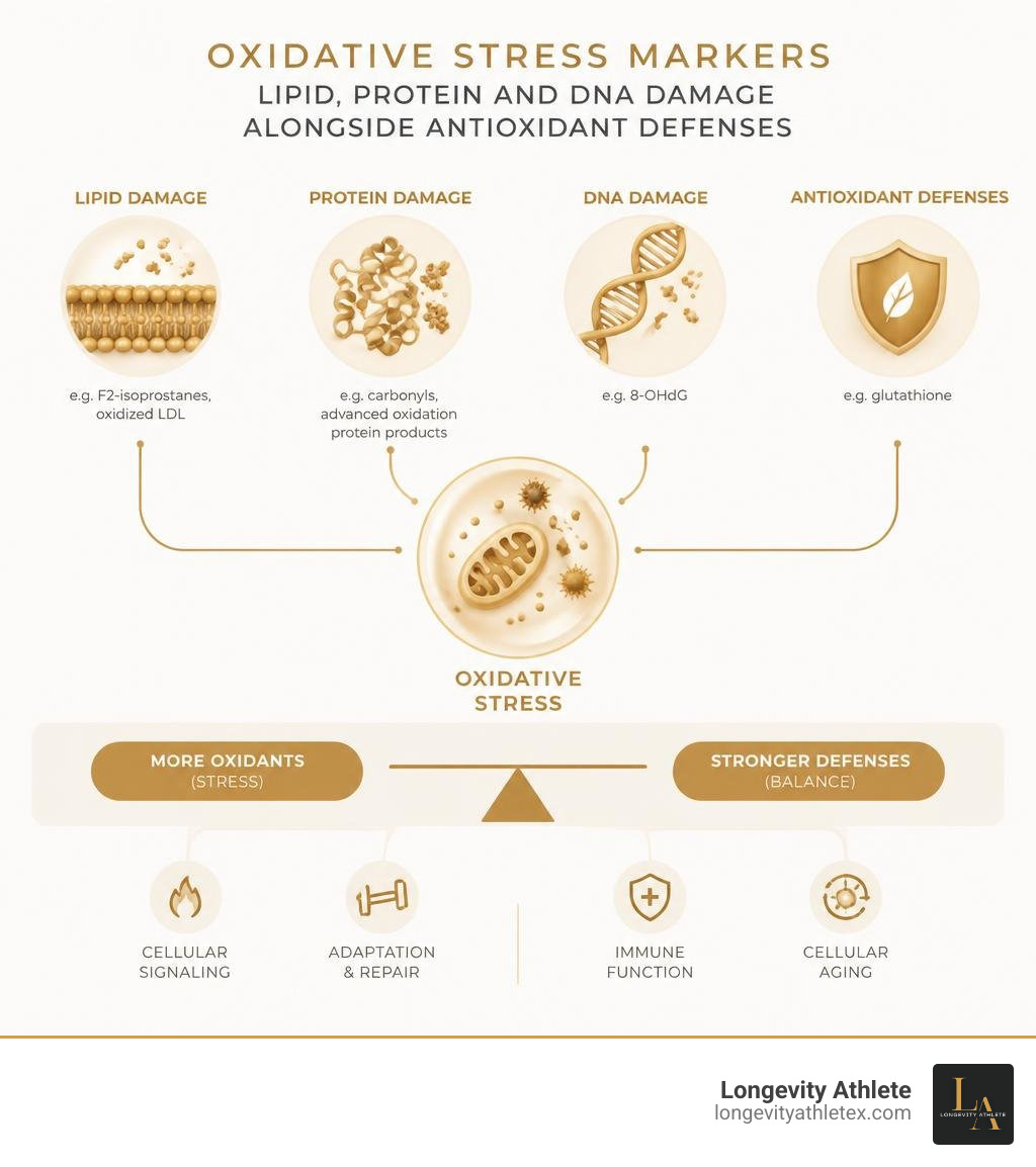
Oxidative stress markers are measurable signs of how the body handles reactive oxygen molecules. Some show damage to fats, proteins, or DNA. Others show how well antioxidant defenses are working. Common examples include F2-isoprostanes, oxidized LDL, 8-OHdG, and glutathione.
These markers can add context to your health and recovery data, but no single result tells the whole story. Levels can shift with exercise, illness, and how a sample is handled. Looking at trends alongside training, sleep, and other health measures is more useful than treating one number as a verdict.
I'm Garric Vosloo. After more than 30 years in patient consulting and years working with elite athletes, I use evidence about oxidative stress markers to help put biomarker results in context.
The Biology of Redox Imbalance: Why Direct ROS Measurement Fails
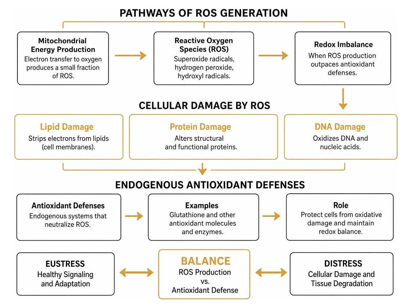
Inside every cell, mitochondria produce energy by transferring electrons to oxygen. During this routine process, a small fraction of oxygen molecules escape complete reduction, generating reactive oxygen species (ROS) such as superoxide radicals, hydrogen peroxide, and hydroxyl radicals. In normal amounts, these molecules serve as vital signaling agents that prompt muscular adaptation, support immune function, and trigger repair pathways.
When the rate of ROS production outpaces the body’s endogenous antioxidant defense capacity, the system shifts from healthy signaling (oxidative eustress) into oxidative distress. Unchecked oxidants begin stripping electrons from lipids, structural proteins, and nucleic acids, contributing to cellular senescence and long-term tissue degradation.
Evaluating this balance directly presents a steep laboratory challenge. Trying to capture reactive oxygen species directly is much like trying to catch a soap bubble before it pops—they vanish almost the instant they appear. Hydroxyl radicals, for instance, possess a half-life measured in nanoseconds and react immediately with whatever cellular structure is closest. Moreover, ROS operate within distinct cellular microdomains, meaning a brief localized burst inside a muscle fiber's mitochondria will not circulate freely through your bloodstream.
Because direct measurements are volatile and impractical, researchers and clinicians rely on stable circulating downstream byproducts. When free radicals damage cell membranes, alter proteins, or oxidize DNA, they leave behind characteristic molecular footprints. A comprehensive review of human redox indexes underscores that tracking these stable chemical modifications provides a reliable, reproducible window into systemic redox balance. Measuring these stable footprints establishes an important biomarker for health and performance evaluation over time.
Key Categories of Oxidative Stress Markers in the Body
Biomarkers of oxidative balance fall into four primary categories based on the physiological substrate modified by free radicals or the enzymatic systems working to neutralize them.
Marker Category | Primary Biomarkers | Analytical Detection Methods | Clinical & Biological Significance |
|---|---|---|---|
Lipid Peroxidation | F2-Isoprostanes, MDA, 4-HNE, OxLDL | LC-MS/MS, GC-MS, HPLC, ELISA, TBARS | Reflects cell membrane degradation, vascular plaque formation, and metabolic load. |
Protein Oxidation | Myeloperoxidase (MPO), Nitrotyrosine, S-Glutathionylation | LC-MS/MS, Immunoassays, Western Blotting | Indicates neutrophil activation, nitric oxide uncoupling, and altered enzyme kinetics. |
DNA / Nucleic Acid Damage | 8-OHdG (8-oxo-dG), 8-oxoG | LC-MS/MS, HPLC-EC, ELISA | Reflects genomic and mitochondrial DNA integrity and oxidative mutation pressure. |
Antioxidant Defenses | SOD, Catalase, Erythrocyte GPX-1, GSH/GSSG Ratio | Spectrophotometry, Enzymatic Assays, HPLC | Measures endogenous capacity to neutralize superoxide, hydrogen peroxide, and lipid peroxides. |
Lipid Peroxidation Byproducts: Isoprostanes, MDA, and OxLDL
Lipids containing polyunsaturated fatty acids (PUFAs) are especially vulnerable to free radical attack due to the reactive hydrogen atoms located between carbon-carbon double bonds. When oxygen radicals attack arachidonic acid, linoleic acid, or docosahexaenoic acid, they trigger a chain reaction that destabilizes cellular membranes.
F2-Isoprostanes (F2-IsoPs): Formed via the non-enzymatic, free-radical-catalyzed peroxidation of arachidonic acid, F2-isoprostanes represent the current gold standard for assessing in vivo lipid peroxidation. Because they are chemically stable and detectable in plasma and urine, they provide an accurate index of systemic oxidative stress. While mass spectrometry (LC-MS/MS or GC-MS) remains the benchmark for precise quantification, commercial ELISA kits offer higher throughput, though they require careful validation against cross-reactive isomers.
Malondialdehyde (MDA): A reactive short-chain aldehyde generated during the breakdown of lipid hydroperoxides. While traditionally measured using the Thiobarbituric Acid Reactive Substances (TBARS) assay, standard TBARS can cross-react with other aldehydes and sugars. High-performance liquid chromatography (HPLC) provides a far more specific measurement. As explored in an analysis of malondialdehyde in inflammatory conditions, MDA levels rise alongside mucosal and systemic inflammatory activity. Furthermore, excessive lipid peroxidation in the central nervous system plays a well-documented role in neurodegenerative processes, as detailed in research exploring the role of lipid peroxidation in neurological disease.
Oxidized Low-Density Lipoprotein (OxLDL): When circulating LDL particles encounter reactive species in the vascular wall, both their lipid core and apolipoprotein B-100 components undergo oxidative modification. OxLDL is recognized by scavenger receptors on macrophages, driving foam cell formation and arterial plaque instability.
Protein Oxidation and Enzymatic Damage: MPO, Nitrotyrosine, and S-Glutathionylation
Proteins carry out vital structural, enzymatic, and transport roles. When ROS or reactive nitrogen species (RNS) oxidize specific amino acid residues, they can disrupt tertiary protein structure, alter binding affinities, or permanently deactivate enzymes.
Myeloperoxidase (MPO): An enzymatic catalyst abundantly expressed in activated neutrophils and monocytes. Upon release during vascular inflammation, MPO uses hydrogen peroxide and chloride ions to generate hypochlorous acid (bleach), while also oxidizing tyrosine residues into 3-chlorotyrosine and 3-nitrotyrosine. Elevated systemic MPO serves as both a functional driver and an early warning marker of vascular lining distress.
Nitrotyrosine: Formed when the potent oxidant peroxynitrite (produced when superoxide reacts rapidly with nitric oxide) nitrates protein-bound tyrosine residues. Elevated 3-nitrotyrosine levels directly reflect nitric oxide consumption and endothelial dysfunction.
S-Glutathionylation: A reversible post-translational modification where a glutathione molecule attaches to reactive cysteine residues on functional proteins, such as the sarco/endoplasmic reticulum Ca2+-ATPase (SERCA). While initially serving as a protective mechanism against irreversible oxidative damage, prolonged S-glutathionylation impairs calcium handling in cardiac and skeletal muscle. Reviewing our comprehensive guide to inflammation markers shows how these oxidative modifications operate hand-in-hand with systemic inflammatory signaling.
DNA and Nucleic Acid Oxidative Stress Markers (8-OHdG)
Nuclear and mitochondrial genomes are susceptible to oxidative modifications, with guanine being the most vulnerable base due to its low oxidation potential. Hydroxyl radical attack at the C-8 position of deoxyguanosine generates 8-hydroxy-2'-deoxyguanosine (8-OHdG), also termed 8-oxo-dG.
When cellular base-excision repair machinery excises this lesion to prevent transversion mutations during replication, the oxidized nucleoside is released into the bloodstream and cleared through the kidneys. Urinary and serum 8-OHdG measurements provide a stable indicator of total-body nucleic acid oxidation. A meta-analysis of F2-isoprostanes and 8-OHdG across chronic disorders observed that 8-OHdG consistently rises across metabolic and neurodegenerative conditions, reflecting cumulative genomic stress.
Endogenous Antioxidant Defense Systems: SOD, Catalase, and Glutathione
Evaluating oxidative status requires looking at both oxidative damage and the enzymatic and non-enzymatic defenses that keep ROS in check:
Superoxide Dismutase (SOD): Enzymes that rapidly convert reactive superoxide radicals into less aggressive hydrogen peroxide and molecular oxygen. Copper-Zinc SOD (SOD1) operates in the cytoplasm, while Manganese SOD (SOD2) protects the mitochondrial matrix.
Catalase (CAT): An enzyme located predominantly in peroxisomes that converts high concentrations of hydrogen peroxide into water and oxygen, protecting cells from peroxide overflow.
Glutathione Peroxidase (GPx) and the GSH/GSSG Ratio: Glutathione (GSH) is the primary intracellular non-protein thiol. GPx enzymes, including erythrocyte GPX-1, utilize reduced GSH to neutralize hydrogen peroxide and lipid hydroperoxides, generating oxidized glutathione disulfide (GSSG). A decreasing ratio of reduced to oxidized glutathione (GSH/GSSG) serves as an intracellular indicator of oxidative shift.
Clinical Significance: Cardiovascular Risk, Metabolic Health, and Disease Progression
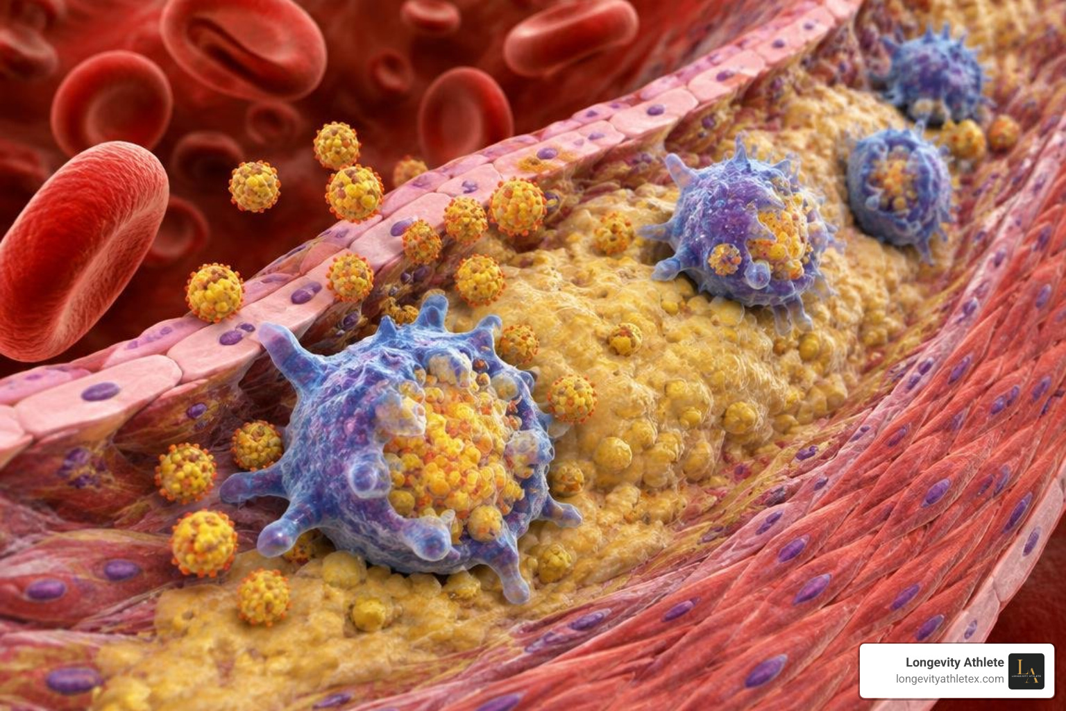
Chronic, unresolved oxidative stress is an established driver in the development of cardiometabolic and vascular conditions.
Vascular Vulnerability and Atherosclerosis
In cardiovascular pathophysiology, oxidative modifications actively drive structural damage within the vessel wall rather than simply signaling its presence:
Plaque Formation and Instability: Elevated plasma protein-bound nitrotyrosine levels are observed in patients with coronary artery disease (CAD), remaining associated with cardiovascular risk even after adjusting for traditional risk factors and C-reactive protein.
Predictive Value of Lipid Peroxides: Research indicates that serum levels of TBARS predicted major cardiovascular events and the need for major vascular procedures over a 3-year follow-up period independently of traditional risk factors in patients with documented coronary artery disease. Furthermore, elevated baseline TBARS predicted carotid atherosclerotic plaque progression over 3 years as assessed by ultrasound wall thickness.
Myeloperoxidase as a Prognostic Signal: Prospective studies demonstrate that elevated circulating MPO levels predict an increased risk of developing CAD in healthy individuals, forecast adverse events in patients presenting with acute chest pain, and track higher risk of myocardial infarction in acute coronary syndrome.
OxLDL Dynamics: Circulating OxLDL levels correlate with clinical disease severity, showing progressive elevations from stable angina to unstable coronary syndromes and acute myocardial infarction, while also serving as an independent predictor of future CAD events in asymptomatic men.
Pairing oxidative metrics with standard laboratory testing, such as testing inflammatory blood markers, provides a clearer picture of vascular health.
Metabolic Dysfunction and Mitochondrial Overload
In skeletal muscle and adipose tissue, chronic nutrient excess creates an oversupply of electrons to complexes I and III of the mitochondrial electron transport chain. As highlighted in research on mitochondrial dysfunction in cardiometabolic diseases, this electron leakage generates excessive superoxide, initiating a cascade of metabolic disruptions:
Impaired Insulin Signaling: Elevated intracellular ROS activates stress-sensitive serine/threonine kinases (such as JNK and IKK-β), which phosphorylate insulin receptor substrate-1 (IRS-1) and block downstream GLUT4 glucose transporter translocation to the cell membrane.
Enzymatic Suppression: Studies have observed reduced superoxide dismutase activity in individuals with obesity, while malondialdehyde markers tend to be higher in individuals with type 2 diabetes compared with healthy controls.
Hemoglobin Modification: Protein modifications such as the S-glutathionylation of hemoglobin are elevated in patients with diabetes, hyperlipidemia, and renal impairment, reflecting ongoing intracellular redox changes.
Circulating Cell-Free mtDNA: Damaged, uncoupled mitochondria release oxidized mitochondrial DNA fragments into the bloodstream, where they can interact with pattern-recognition receptors to amplify systemic inflammatory responses.
Practical Applications: Using Redox Profiling to Guide Nutrition and Training

For athletes, high performers, and longevity-focused individuals, managing oxidative stress requires a balanced approach. Complete suppression of free radicals is counterproductive, as exercise-induced ROS are necessary physiological signals for adaptation.
Balancing Exercise Hormesis and Preventing Training Blunting
Intense muscular contraction generates a controlled burst of superoxide and hydrogen peroxide. This transient oxidative pulse activates the peroxisome proliferator-activated receptor gamma coactivator 1-alpha (PGC-1α) and AMP-activated protein kinase (AMPK) pathways, which coordinate mitochondrial biogenesis, improve capillary density, and upregulate endogenous SOD and GPx enzymes.
Taking high-dose, non-selective antioxidant supplements (such as 1,000 mg of vitamin C or high-dose vitamin E) directly before or after training sessions can blunt this adaptive signal, potentially reducing endurance gains and muscular remodeling.
Instead, a practical strategy emphasizes nutrient-dense, whole-food polyphenol sources. A systematic evaluation of dietary berries on oxidative markers found that regular consumption of anthocyanin-rich fruits supported modest reductions in circulating OxLDL and MDA without suppressing exercise adaptations. Structuring dietary antioxidant timing away from strenuous training bouts supports recovery while preserving adaptive signaling, a core principle when designing biomarker-based nutrition plans.
Interpreting Oxidative Stress Markers for Athletic Recovery and Longevity
Evaluating individual redox markers can help tailor training volume and lifestyle strategies:
Establish Stable Baseline Levels: Check markers such as F2-isoprostanes, urinary 8-OHdG, OxLDL, and erythrocyte GPX-1 activity during a baseline training phase. Erythrocyte GPX-1 activity has been shown to correlate inversely with cardiovascular events, highlighting the value of maintaining robust endogenous enzyme reserves.
Monitor High-Intensity Training Cycles: During intense training blocks or endurance preparation, tracking lipid peroxidation byproducts and tracking inflammation and oxidative recovery in athletes can help spot overreaching before it turns into chronic fatigue.
Assess Dietary and Lifestyle Adjustments: If follow-up tests show elevated 8-OHdG or high OxLDL, personalized lifestyle adjustments—such as increasing polyphenol intake, adjusting sleep schedules, or altering training intensity—can be implemented and monitored over 8 to 12 weeks to gauge response.
Frequently Asked Questions About Oxidative Stress Biomarkers
Which biomarker is considered the gold standard for lipid peroxidation?
F2-isoprostanes are widely regarded by researchers as the gold standard for measuring in vivo lipid peroxidation. Because they are formed specifically through the non-enzymatic free-radical attack of arachidonic acid in membrane phospholipids, their quantification via liquid chromatography-tandem mass spectrometry (LC-MS/MS) provides an accurate, reproducible assessment of systemic lipid damage.
Why is direct measurement of reactive oxygen species impractical?
Direct measurement of ROS in blood is impractical because most free radical species possess extremely short half-lives (ranging from nanoseconds to microseconds) and react immediately with adjacent cellular structures. Furthermore, ROS are generated within specific sub-cellular compartments rather than circulating freely, making stable downstream oxidation byproducts (such as F2-isoprostanes, MDA, and 8-OHdG) the preferred markers for clinical and athletic assessment.
Can high-dose antioxidant supplements negatively impact athletic training?
Yes, research suggests that taking high-dose antioxidant supplements (like large daily doses of vitamins C and E) immediately around workout windows may blunt muscular and endurance adaptations. Exercise-induced ROS act as essential signaling molecules that activate PGC-1α and stimulate mitochondrial biogenesis; neutralizing them completely can diminish the body's natural training response.
Personalized Redox Profiling for Longevity
Balancing cellular oxidation and antioxidant defense is central to maintaining long-term metabolic, cardiovascular, and physical health. Rather than guessing with generic supplement stacks, tracking validated oxidative stress markers provides actionable data to refine training loads, optimize recovery windows, and support cellular health.
At Longevity Athlete, we provide precision-performance wellness consulting for disciplined athletes, using personalized biomarker testing and data-driven plans to optimize longevity and physical output.
Our programs are structured to meet your specific goals:
The Signature Plan is our self-guided group program designed to help you understand your core metrics and implement foundational health practices.
Our Gold and Platinum Plans deliver individualized lifestyle programs featuring comprehensive biomarker analysis, custom training calibrations, and ongoing support tailored to your unique biology.
Explore how optimizing performance biomarkers can support your training, protect cellular health, and sustain long-term performance.
Scientific References and Further Reading
Ho E, Karimi Galougahi K, Liu CC, Bhindi R, Figtree GA. "Biological markers of oxidative stress: Applications to cardiovascular research and practice.." Redox biology, 2013. PMCID PMC3830063. Shichiri M. "The role of lipid peroxidation in neurological disorders.." Journal of clinical biochemistry and nutrition, 2014. PMCID PMC4042144. Cordiano R, Di Gioacchino M, Mangifesta R, Panzera C, Gangemi S, Minciullo PL. "Malondialdehyde as a Potential Oxidative Stress Marker for Allergy-Oriented Diseases: An Update.." Molecules (Basel, Switzerland), 2023. PMCID PMC10457993. Msigwa SS, Mkwambe MC, Yu QY, Zhu SG, Zhu JH, Zhang X, Wang JY. "Comparative evaluation of oxidative stress biomarkers F2-isoprostanes and 8-OHdG in Parkinson's disease and Type 2 Diabetes Mellitus: a systematic review and meta-analysis of human studies.." Annals of medicine, 2026. PMCID PMC13097182. Sánchez-Rodríguez MA, Mendoza-Núñez VM. "Oxidative Stress Indexes for Diagnosis of Health or Disease in Humans.." Oxidative medicine and cellular longevity, 2019. PMCID PMC6899293. Stote KS, Burns G, Mears K, Sweeney M, Blanton C. "The Effect of Berry Consumption on Oxidative Stress Biomarkers: A Systematic Review of Randomized Controlled Trials in Humans.." Antioxidants (Basel, Switzerland), 2023. PMCID PMC10376627. Cojocaru KA, Luchian I, Goriuc A, Antoci LM, Ciobanu CG, Popescu R, Vlad CE, Blaj M, Foia LG. "Mitochondrial Dysfunction, Oxidative Stress, and Therapeutic Strategies in Diabetes, Obesity, and Cardiovascular Disease.." Antioxidants (Basel, Switzerland), 2023. PMCID PMC10045078.