Master Gland Mechanics: A Practical Guide to Pituitary Blood Testing
What a Pituitary Gland Function Blood Test Can Tell You
A pituitary gland function blood test checks whether this small gland is sending the right hormone signals. A clinician may measure pituitary hormones, such as ACTH, TSH, LH, FSH, and prolactin, alongside hormones from the glands they control. The results can help explain symptoms such as persistent fatigue, changes in menstrual cycles, or low sex hormone levels. One blood draw may not tell the whole story: timing matters, and some results call for further testing.
The pituitary is called the master gland because it helps direct growth, thyroid function, stress response, and reproduction. This guide explains what the tests measure, how to prepare, and why a clinician might recommend follow-up tests or an MRI.
I’m Garric Vosloo. In more than 30 years of health consulting, including work with elite athletes, I’ve learned why a pituitary gland function blood test needs to be understood alongside symptoms and other health data.
Understanding the Master Gland: Physiology and Systemic Regulation
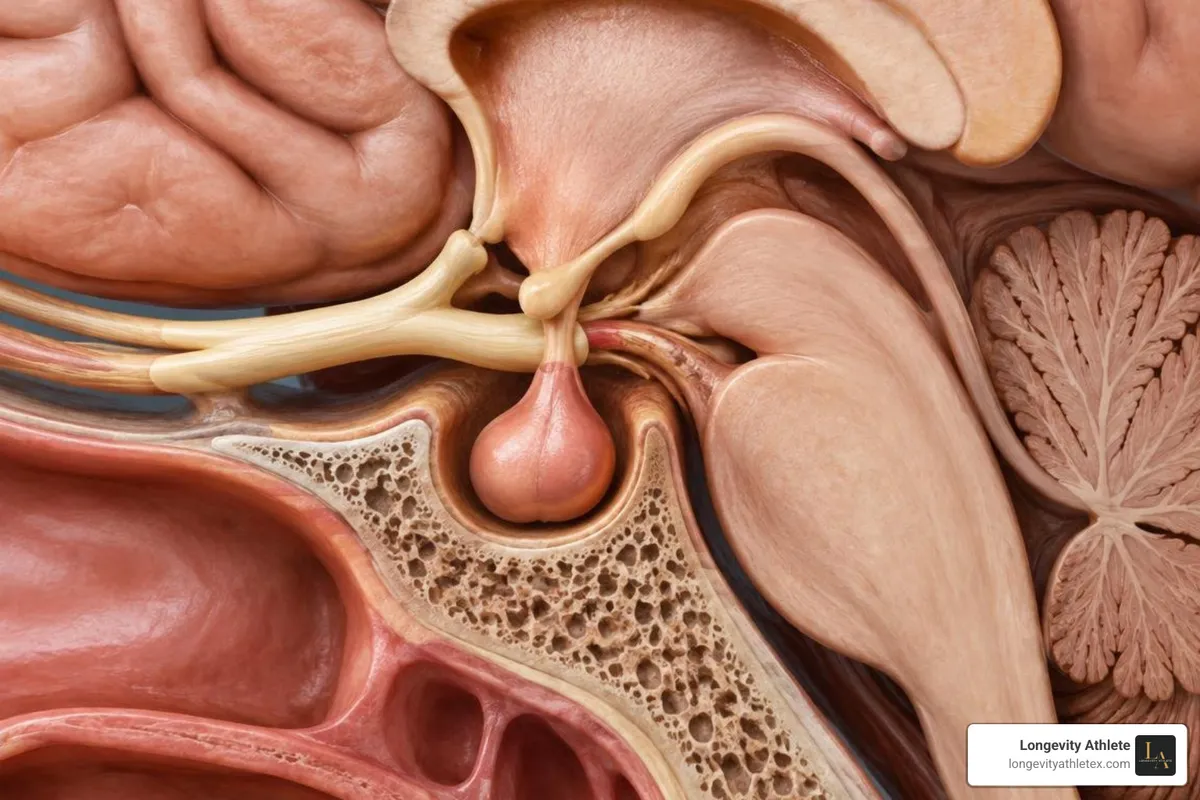
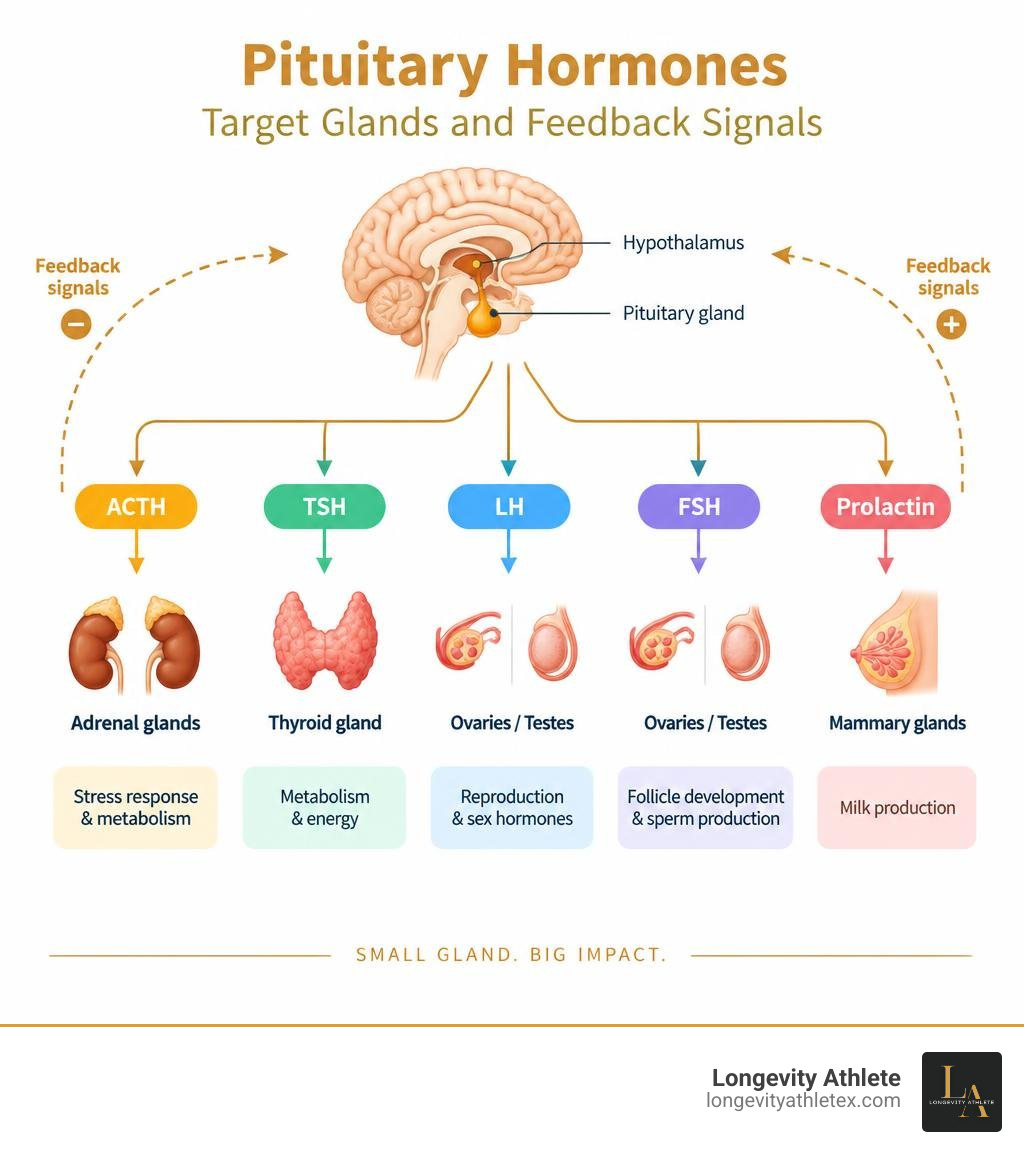
Nestled securely within the bony cradle of the sphenoid bone known as the sella turcica, the pituitary gland serves as the primary operational hub of the endocrine system. Despite being roughly the size of a pea, it manages the chemical messages that govern systemic vitality, basal metabolic rate, and cellular repair. It does not act in isolation; rather, it operates under the direct supervision of the hypothalamus, which monitors systemic parameters and releases stimulating or inhibiting neurohormones to direct pituitary secretion.
This communication relies on tight negative feedback loops. When circulating levels of target organ hormones (such as cortisol, free thyroxine, or testosterone) reach an optimal threshold, they signal both the hypothalamus and the pituitary to reduce trophic hormone output. When peripheral levels drop, the pituitary responds by increasing its signaling hormones. In the clinical evaluation of systemic wellness and an overview of endocrine disorders, distinguishing whether a hormonal deficiency originates within the target gland (primary dysfunction) or within the pituitary/hypothalamus (secondary or central dysfunction) forms the cornerstone of accurate diagnostic work.
Anterior vs. Posterior Pituitary Hormones
The pituitary gland is anatomically and functionally divided into two distinct lobes: the anterior pituitary (adenohypophysis) and the posterior pituitary (neurohypophysis).
The Anterior Pituitary: Composed of glandular tissue, the anterior lobe synthesizes and secretes six major trophic hormones into the systemic circulation:
Adrenocorticotropic Hormone (ACTH) – directs the adrenal cortex
Thyroid-Stimulating Hormone (TSH) – stimulates the thyroid gland
Luteinizing Hormone (LH) & Follicle-Stimulating Hormone (FSH) – orchestrate gonadal sex steroid production and gametogenesis
Growth Hormone (GH) – drives tissue growth, protein synthesis, and cellular repair
Prolactin (PRL) – governs lactation and influences reproductive behavior
The Posterior Pituitary: An extension of neural tissue directly connected to the hypothalamus, this lobe does not synthesize its own hormones. Instead, it stores and releases two hypothalamic peptide hormones:
Arginine Vasopressin (AVP / Antidiuretic Hormone) – regulates renal water reabsorption and plasma osmolality
Oxytocin – manages uterine contractions, milk ejection, and key aspects of social bonding
Understanding the specific biological behavior of each hormone makes it easier to select the right important biomarker for health and performance when mapping systemic adaptation.
Clinical Indicators for a Pituitary Gland Function Blood Test
Because the pituitary gland coordinates multiple organ systems, clinical signs of dysfunction can vary widely. A practitioner may evaluate pituitary function when an individual presents with subtle, multisystem complaints that do not match isolated end-organ pathology.
Common clinical indicators include:
Unexplained chronic fatigue and exercise intolerance that fails to resolve with sleep
Visual field deficits (specifically bitemporal hemianopsia caused by optic chiasm compression from sellar enlargement)
Persistent headaches combined with systemic energy drops
Galactorrhea (unexplained breast milk production) or marked hyperprolactinemia symptoms
Secondary amenorrhea or oligomenorrhea in women without anatomical pelvic abnormalities
Unexplained low morning testosterone, erectile dysfunction, or loss of libido in men
Orthostatic hypotension, severe dizziness, and electrolyte imbalances (such as hyponatremia)
Unintended, rapid fluctuations in body weight or loss of lean mass despite stable nutrition
For high-performing individuals, tracking these signals helps protect foundational performance biomarkers before deep systemic fatigue sets in.
Core Biomarkers in a Pituitary Gland Function Blood Test Panel
A comprehensive baseline assessment requires measuring the pituitary's trophic hormones alongside the peripheral hormones produced by their target glands. Looking at a pituitary hormone in isolation often provides an incomplete picture. For example, a "normal" TSH value in the presence of an abnormally low free thyroxine level points directly to central, secondary hypopituitarism rather than healthy thyroid regulation.
Pituitary Hormone | Target Organ / Axis | Optimal Draw Window | Typical Clinical Deficiency Sign | Typical Clinical Excess Sign |
|---|---|---|---|---|
ACTH | Adrenal Cortex | 08:00 AM (fasting) | Fatigue, hypotension, weight loss | Cushing's disease, central adiposity |
TSH | Thyroid Gland | Early morning | Secondary hypothyroidism, lethargy | Secondary hyperthyroidism, tachycardia |
LH / FSH | Gonads (Ovaries/Testes) | Morning (Day 2–4 of cycle in women) | Secondary hypogonadism, amenorrhea | Premature puberty, rare gonadal adenomas |
Growth Hormone (GH) | Systemic / Liver | Morning fasting (pulsatile) | Decreased recovery, sarcopenia | Gigantism (youth), Acromegaly (adult) |
IGF-1 | Liver (GH proxy) | Any time (stable half-life) | Suboptimal tissue repair, low vitality | Acromegaly, soft tissue overgrowth |
Prolactin | Mammary Gland / Gonads | Resting morning, fasting | Inability to lactate postpartum | Galactorrhea, hypogonadism, infertility |
Adrenocortical and Thyroid Axis Markers (ACTH, Cortisol, TSH, Free T4)
Evaluating the adrenocortical axis requires synchronized measurement of plasma ACTH and serum total cortisol. Cortisol production follows a robust circadian rhythm, reaching peak circulating concentrations between 07:00 AM and 09:00 AM before tapering toward a nadir around midnight. A morning serum cortisol draw below reference thresholds alongside a low or inappropriately "normal" plasma ACTH strongly suggests secondary (central) adrenal insufficiency.
Similarly, evaluating the central thyroid axis requires pairing TSH with free thyroxine (Free T4). In classic primary hypothyroidism, a failing thyroid produces low Free T4, prompting the pituitary to elevate TSH. However, in central hypothyroidism, the pituitary fails to produce sufficient TSH; consequently, blood tests show low Free T4 levels accompanied by a normal or low TSH. Relying solely on a TSH screening test risks missing this central failure entirely.
Somatotropic and Gonadal Axis Markers (GH, IGF-1, Prolactin, LH, FSH)
Evaluating growth hormone status presents a unique laboratory challenge because the anterior pituitary releases GH in brief pulses rather than a steady stream. A random resting blood draw for growth hormone is often of limited diagnostic value because physiological levels frequently dip below detection limits between secretory pulses. For this reason, clinicians measure Insulin-like Growth Factor 1 (IGF-1) as a dependable proxy. Synthesized primarily in the liver under growth hormone stimulation, IGF-1 circulates bound to transport proteins with a stable half-life, providing an integrated reflection of daily growth hormone output.
Prolactin assessment requires careful resting conditions, as physical stress, venipuncture anxiety, and recent exercise can transiently elevate serum concentrations. When significantly elevated, labs may also run dilution assays or polyethylene glycol (PEG) precipitation to rule out assay artifacts like the "hook effect" or the presence of macroprolactin.
Finally, the gonadal axis relies on matching Luteinizing Hormone (LH) and Follicle-Stimulating Hormone (FSH) against circulating sex steroids—total and free testosterone in men, and estradiol and progesterone in women. In individuals undergoing routine sports bloodwork, low systemic testosterone or estradiol accompanied by low or un-elevated LH/FSH indicates secondary hypogonadism, which prompts further investigation of the pituitary-hypothalamic connection.
Dynamic Endocrine Testing vs. Basal Bloodwork
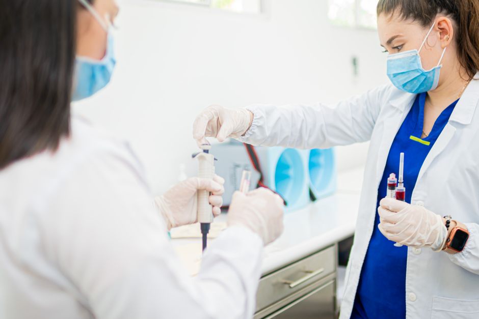
While resting baseline blood tests provide valuable clues, static measurements cannot always confirm whether a gland has adequate functional reserve or the ability to turn off when prompted. In these borderline scenarios, clinicians use dynamic endocrine testing. Dynamic evaluation involves introducing a pharmacological stimulus to see if the gland can respond appropriately (a stimulation test for suspected deficiency), or administering a suppressive compound to check if overactive production can be quieted (a suppression test for suspected autonomous hypersecretion).
Stimulation Assays: Insulin Tolerance, Synacthen, and Arginine Tests
When basic morning labs suggest hypopituitarism, stimulation tests evaluate the pituitary's capacity to respond to physiological stress:
Insulin Tolerance Test (ITT): Regarded as a gold standard for evaluating both ACTH and growth hormone reserves. Intravenous regular insulin is administered under close clinical supervision to induce transient hypoglycemia (blood glucose reaching 2.2 mmol/L or lower). Hypoglycemia serves as a potent central stressor that should prompt an immediate surge in both ACTH (driving serum cortisol above 500 nmol/L) and growth hormone.
Synacthen (Cosyntropin) Test: Involves administering synthetic ACTH to assess adrenocortical responsiveness. While primarily evaluating adrenal capacity, an inadequate rise in individuals with longstanding central ACTH deficiency reflects secondary adrenal atrophy from lack of chronic pituitary signaling.
Arginine Infusion & Glucagon Stimulation Tests: Used when an insulin tolerance test is clinically contraindicated (such as in individuals with cardiovascular conditions or seizure disorders). These agents trigger central pathways to stimulate growth hormone release, allowing clinicians to verify true somatotroph deficiency through dedicated growth hormone testing.
Suppression Assays and Posterior Pituitary Osmolality Evaluation
When clinicians suspect autonomous, uncontrolled hormone overproduction (such as from a functional pituitary microadenoma), suppression testing is required:
Dexamethasone Suppression Testing: An individual takes a synthetic glucocorticoid (dexamethasone) late in the evening, and serum cortisol is measured the following morning at 08:00 AM. In a healthy pituitary-adrenal feedback loop, dexamethasone signals the pituitary to suppress ACTH, driving morning cortisol down. Failure to suppress suggests autonomous ACTH secretion (Cushing's disease) or direct adrenal hypersecretion.
Oral Glucose Tolerance Test (OGTT) for Growth Hormone: In healthy physiology, a high-carbohydrate glucose load suppresses growth hormone secretion to near-undetectable levels. In individuals with acromegaly, growth hormone levels remain unsuppressed, confirming autonomous secretion.
Water Deprivation Testing: Used to evaluate posterior pituitary function and antidiuretic hormone release. When an individual is restricted from fluids under structured supervision, serum and urine osmolality are tracked hourly. If the posterior pituitary fails to release vasopressin despite rising serum concentration, urine remains inappropriately dilute, pointing to central diabetes insipidus rather than nephrogenic causes.
Diagnostic Preparation and Sellar Neuroimaging
Pre-analytical accuracy is critical when collecting samples for an endocrine panel. Because pituitary hormones are sensitive to circadian rhythms, emotional stress, metabolic states, and dietary supplements, proper preparation helps prevent misleading test results.
How to Prepare for a Blood Test for Pituitary Gland Function
To ensure the blood sample reflects true baseline physiology, clinicians generally recommend several specific preparation steps:
Observe Overnight Fasting: Fast for 8 to 12 hours before morning blood collection. Avoid caloric intake while staying well-hydrated with plain water.
Standardize Collection Timing: Schedule your appointment between 07:30 AM and 09:00 AM. This early window is essential for accurate ACTH, morning cortisol, and testosterone assessments, all of which peak early in the day.
Complete a Biotin Washout: Pause high-dose biotin (vitamin B7) supplements for 7 to 10 days before blood collection. Biotin can interfere with common laboratory immunoassays, leading to falsely elevated or falsely suppressed results across multiple hormone tests.
Coordinate Menstrual Cycle Timing: For reproductive-age women not on hormonal birth control, baseline gonadotropins (LH, FSH) and estradiol are typically drawn during the early follicular phase (Days 2 to 4 of the menstrual cycle) unless directed otherwise.
Minimize Acute Morning Stress: Arrive at the laboratory early and rest quietly for 15 to 20 minutes before the blood draw. Physical exertion or rushing can artificially elevate prolactin and ACTH levels.
Sellar Magnetic Resonance Imaging (MRI)
When blood tests indicate pituitary hormone excess or central deficiency, imaging provides high-resolution anatomical context. Dedicated sellar Magnetic Resonance Imaging (MRI)—performed with thin-slice T1- and T2-weighted sequences both before and after gadolinium contrast administration—serves as the primary imaging modality for evaluating sellar and parasellar structures.
Neuroimaging can identify several key structural findings:
Microadenomas: Benign tumors measuring less than 10 millimeters in diameter, often responsible for functional hypersecretion (such as prolactinomas or ACTH-secreting lesions).
Macroadenomas: Tumors measuring 10 millimeters or larger. These lesions can cause hormone overproduction or create "mass effect" compression on neighboring healthy pituitary cells, leading to hypopituitarism. They may also compress the optic chiasm, causing visual field changes.
Empty Sella Syndrome: A condition where the subarachnoid space herniates into the sella turcica, flattening the pituitary tissue against the bony floor.
Infiltrative or Ischemic Changes: Evidence of hypophysitis, vascular injury, or localized tissue loss.
Differential Diagnosis and Clinical Management Pathways
Interpreting pituitary blood tests requires looking at all hormone axes together. Because multiple hormone signals originate from a single anatomical gland, a structural or functional disruption in one area often affects neighboring pathways.
Differentiating Functional Adenomas from Hypopituitarism
Clinical findings generally fall into two broad categories: functional adenomas that actively overproduce specific hormones, or tissue damage that leads to progressive hypopituitarism.
Prolactinomas: The most common functional pituitary tumor. Markedly elevated baseline prolactin accompanied by low LH/FSH often responds well to medical therapy with dopamine agonists (such as cabergoline), which help reduce tumor size and normalize hormone levels.
Cushing's Disease: Characterized by autonomous ACTH secretion driving sustained cortisol excess. Management typically involves transsphenoidal surgical resection of the microadenoma.
Acromegaly: Driven by excessive growth hormone and IGF-1 production, causing soft tissue expansion and metabolic shifts. Treatment often combines surgical management with medical therapies like somatostatin analogs.
Nonfunctional Adenomas & Mass-Effect Hypopituitarism: These tumors do not secrete active hormones but can compress healthy pituitary tissue as they grow. This compression can lead to sequential hormone decline, typically affecting growth hormone and gonadotropins first, followed by TSH and ACTH. In these situations, individuals often benefit from personalized biomarker analysis to monitor hormone balance and guide replacement therapies.
Postpartum Necrosis and Sheehan's Syndrome Workup
A distinct form of secondary hypopituitarism is Sheehan's syndrome, which develops following severe obstetric hemorrhage and hypotension during childbirth. During pregnancy, the anterior pituitary enlarges significantly, increasing its metabolic demand and vulnerability to ischemic injury if systemic perfusion drops sharply.
Classic early indicators include an inability to produce breast milk (agalactia from acute prolactin deficiency) alongside persistent postpartum fatigue and a failure to resume normal menstrual cycles. Diagnostic blood tests reveal low circulating levels across multiple anterior pituitary axes (frequently involving GH, gonadotropins, ACTH, and TSH). In chronic cases, sellar MRI typically shows pituitary atrophy or an empty sella. Clinical management focuses on structured, lifelong hormone replacement, prioritizing adrenal and thyroid support before addressing sex steroids and growth hormone.
Frequently Asked Questions About Pituitary Gland Function Testing
What is the most common reason a doctor orders a pituitary blood test?
Clinicians most frequently request pituitary blood tests to evaluate unexplained chronic fatigue, central metabolic issues, and reproductive challenges. Common triggers include low morning testosterone with normal or low gonadotropins in men, secondary amenorrhea in women, persistent galactorrhea, or thyroid hormone patterns that do not fit classic primary thyroid disease. They are also standard when follow-up is needed for incidental sellar findings on brain imaging.
Why is random growth hormone testing often insufficient for diagnosis?
The anterior pituitary releases growth hormone in brief, pulsatile bursts throughout the day and night, with levels dipping to near-zero between peaks. A single, random blood draw cannot reliably distinguish a normal resting trough from true growth hormone deficiency. To get a clearer clinical picture, practitioners evaluate serum IGF-1 as a stable proxy and use formal dynamic stimulation testing when assessing overall growth hormone reserve.
How long should I stop biotin before undergoing pituitary blood tests?
Most clinical laboratories recommend stopping high-dose biotin (vitamin B7) supplements for 7 to 10 days before an endocrine blood draw. Biotin is widely used in high-throughput immunoassay testing platforms. Elevated levels of circulating biotin in the blood sample can interfere with test reagents, causing false-positive elevations or misleading suppressions across thyroid, adrenal, and reproductive hormone panels.
Conclusion
The pituitary gland plays an indispensable role in maintaining systemic balance, athletic recovery, and metabolic health. Because endocrine pathways interact through complex feedback loops, evaluating these signals requires careful timing, proper pre-test preparation, and a combined look at both trophic and end-organ markers.
At Longevity Athlete, we emphasize precision tracking to help high-performing individuals understand their unique hormonal data. Our Signature Plan provides a self-guided group experience for foundational wellness education, while our Gold and Platinum Plans offer individualized lifestyle plans built around comprehensive testing. If you are experiencing unexplained recovery challenges or want to establish an accurate health baseline, working with qualified clinical professionals to evaluate your hormone panel provides a reliable foundation for long-term health and performance.